Review
. 2012 Jul;60(7):1013-23.
doi: 10.1002/glia.22288. Epub 2012 Jan 6.

Artifact versus reality--how astrocytes contribute to synaptic events

Affiliations
Review

Artifact versus reality--how astrocytes contribute to synaptic events

Maiken Nedergaard et al. Glia. 2012 Jul.

Abstract

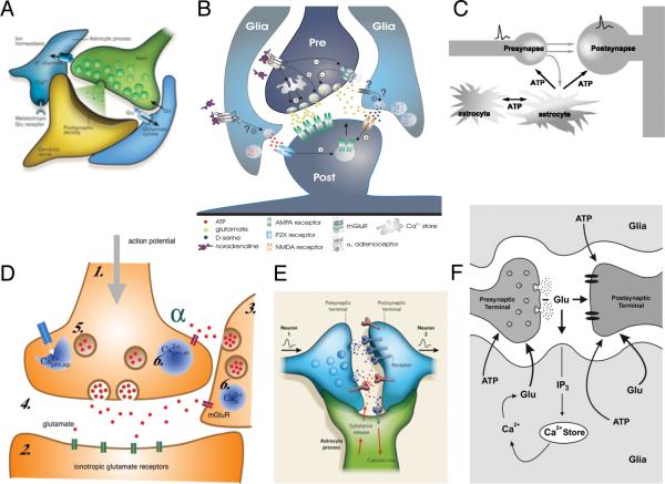
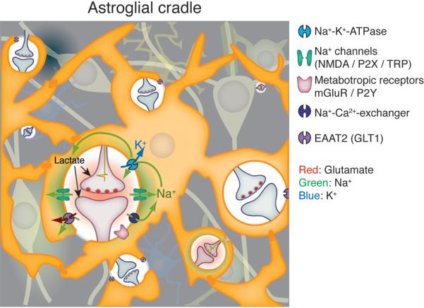
The neuronal doctrine, developed a century ago regards neuronal networks as the sole substrate of higher brain function. Recent advances in glial physiology have promoted an alternative hypothesis, which places information processing in the brain into integrated neuronal-glial networks utilizing both binary (neuronal action potentials) and analogue (diffusional propagation of second messengers/metabolites through gap junctions or transmitters through the interstitial space) signal encoding. It has been proposed that the feed-forward and feed-back communication between these two types of neural cells, which underlies information transfer and processing, is accomplished by the release of neurotransmitters from neuronal terminals as well as from astroglial processes. Understanding of this subject, however, remains incomplete and important questions and controversies require resolution. Here we propose that the primary function of perisynaptic glial processes is to create an "astroglial cradle" that shields the synapse from a multitude of extrasynaptic signaling events and provides for multifaceted support and long-term plasticity of synaptic contacts through variety of mechanisms, which may not necessarily involve the release of "glio" transmitters.


Figures

Figure 1
Figure 1. The tripartite synapse
The model of the tripartite synapse is based on the observation that glutamate or GABA trigger Ca2+ increases in peri-synaptic glial processes in an mGlu1/5 receptor-dependent pathway (or by GABAB receptors in inhibitory synapses). In turn, the increase in astrocytic Ca2+ triggers vesicular gliotransmitters release, which modulates synaptic strength by binding to pre- and postsynaptic receptors. Six recent diagrams of the tripartite synapse illustrating that the tripartite synapse is viewed as the epicenter for neuron-glia signaling. A (Eroglu and Barres, 2010), B (Gordon and Bains 2006), C (Koizumi et al. 2005), D (Nadkarni et al. 2008) E (Allen and Barres 2009), F (Sotero and Martinez-Cancino 2010).
Figure 2
Figure 2. Ca2+ signaling in cultured astrocytes trigger release of multiple amino acids, not only glutamate
(A) Amino acids in the cytosol of cultured rat astrocytes. (B) Amino acid release in response to exposure to a hypotonic solution (214 mOsm). (C) Amino acid release in response to the purinergic receptor agonist ATP (100 μM). Glutamate is not released in isolation, but rather together with other amino acids present in the cytosol of astrocytes. Modified from (Takano et al. 2005).
Figure 3
Figure 3. The astroglial cradle
In the “astroglial cradle” model, the main function of peri-synaptic glial processes is to isolate and support the function of individual synapses, thus ensuring the spatial specificity of synaptic transmissions. The physical isolation of synapses first noted by Ramon y Cajal is complemented by transporters (GLT1 and other), which together provide a functional barrier isolating the synapse from signaling events in the extrasynaptic space. The astroglial membranes contain several types of ionotropic receptors, including NMDA and P2X receptors located close to the sites of neurotransmitter release from presynaptic terminals (Lalo et al. 2006; Lalo et al. 2008). Although these receptors are functionally similar to those expressed by neurons, ionotropic receptors assume a different function in the electrically non-excitable membranes of astrocytes (indeed, the amplitudes of synaptically induced ionotropic receptor-mediated currents measured from astrocytes rarely exceed 50 pA, whereas typical input resistance of astrocytes in situ is ~ 50 mOhm (Lalo et al. 2006); the resulting depolarization cannot, therefore, exceed 2 - 3 mV). Activation of astrocytic NMDA and P2X receptors generate inward cationic (mainly carried by Na+) currents, which rapidly increase cytosolic Na+ concentration in perisynaptic astrocytic processes. The transient increase of [Na+]I, has several functional consequences: (1) The increase in [Na+]i forces the reverse mode of Na+/Ca2+ exchanger (NCX; the latter is strategically co-localized with glutamate transporters in perisynaptic astroglial processes) (Minelli et al. 2007), which produces rapid local Ca2+ increases in the perisynaptic processes of active synapses. (2) The decrease in transmembrane Na+ gradient transiently reduces the efficacy of Na+-dependent glutamate transport, thereby slowing down elimination of synaptically released glutamate. (Kirischuk et al. 1997). (3) The excess of [Na+]i activates Na+/K+ ATPase, which in turn decreases extracellular K+ and enhances lactate production. The latter provides focal metabolic support to active synapses (Pellerin et al. 2007). In addition, elevation in [Ca2+]i (due to activation of metabotropic receptros with subsequent Ca2+ mobilization from the stores or due to plasmalemmal Ca2+ entry) directly enhances the activity of the Na+/K+ ATPase providing a longer lasting decrease in extracellular K+ and further maintenance of lactate shuttle. Combined activity of Na+/K+ pump and NCX eventually lower [Na+]i thus restoring glutamate accumulation. In summary, activation of ionotropic receptors ion transporters and exchngers in astroglia membranes initiates fast neuronal-glial-neuronal interactions (msec) at the level of the individual synapse, whereas Ca2+ signaling provides for longer lasting modulation (seconds). Combined, the signaling events evoked by activation of astrocytic ionotropic and metabotropic receptors enable astrocytes to modulate synaptic transmission on multiple levels, including control of the peak amplitude and duration of glutamate increase, extracellular K+ concentration, and energy substrate delivery (lactate) employing pathways that do not involve gliotransmitter release.
Figure 4
Figure 4. Morphological plasticity of astrocytes
(A) Golgi-stained astrocytes from a 2 month-old human infant in cortical layers I-III. A, B, C, and D are cells in layer I, whereas E,F,G and H are astrocytes in layer II and II. I and J are cells with endfeet contacting blood vessels. V, blood vessel. (Ramón y Cajal, 1913). (B) Cultured rat astrocytes before (top panel) and after addition of dBcAMP (1 μM, lower panel), cresyl violet. Courtesy of Drs. Fujita and Abe, Hoshi University. (C) Electron microscopy of the rat SON during parturition (top panel) and lactation (lower panel). Astrocytes withdraw their processes in lactating animals. From (Theodosis et al. 2008).

References

    1. Adachi K, Cruz NF, Sokoloff L, Dienel GA. Labeling of metabolic pools by [6-14C]glucose during K+-induced stimulation of glucose utilization in rat brain. Journal of cerebral blood flow and metabolism : official journal of the International Society of Cerebral Blood Flow and Metabolism. 1995;15:97–110. - PubMed
    1. Agulhon C, Fiacco TA, McCarthy KD. Hippocampal short- and long-term plasticity are not modulated by astrocyte Ca2+ signaling. Science. 2010;327:1250–4. - PubMed
    1. Agulhon C, Petravicz J, McMullen AB, Sweger EJ, Minton SK, Taves SR, Casper KB, Fiacco TA, McCarthy KD. What is the role of astrocyte calcium in neurophysiology? Neuron. 2008;59:932–46. - PMC - PubMed
    1. Airan RD, Thompson KR, Fenno LE, Bernstein H, Deisseroth K. Temporally precise in vivo control of intracellular signalling. Nature. 2009;458:1025–9. - PubMed
    1. Allen NJ, Barres BA. Neuroscience: Glia - more than just brain glue. Nature. 2009;457:675–7. - PubMed

Publication types