. 2012 Feb 1;18(3):654-65.
doi: 10.1158/1078-0432.CCR-11-1406. Epub 2012 Jan 6.

Notch signaling promotes growth and invasion in uveal melanoma

Affiliations

Notch signaling promotes growth and invasion in uveal melanoma

Laura Asnaghi et al. Clin Cancer Res. .

Abstract

Purpose: To determine whether uveal melanoma, the most common primary intraocular malignancy in adults, requires Notch activity for growth and metastasis.

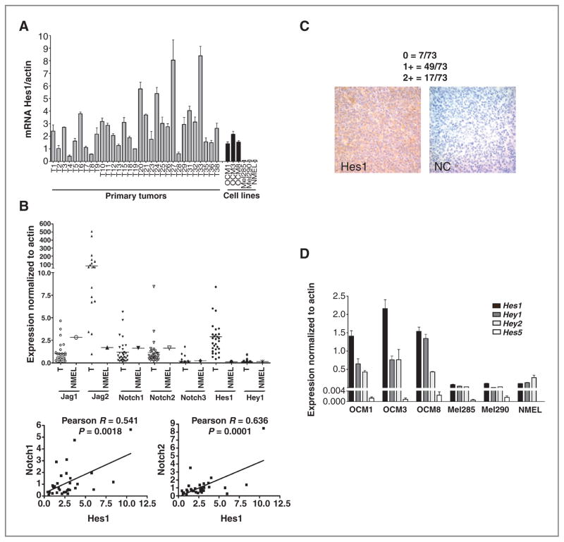
Experimental design: Expression of Notch pathway members was characterized in primary tumor samples and in cell lines, along with the effects of Notch inhibition or activation on tumor growth and invasion.

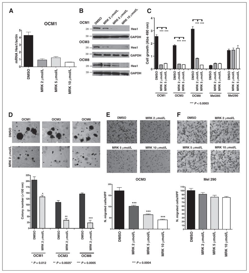
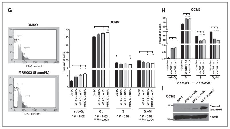
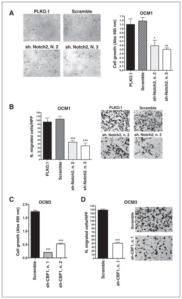
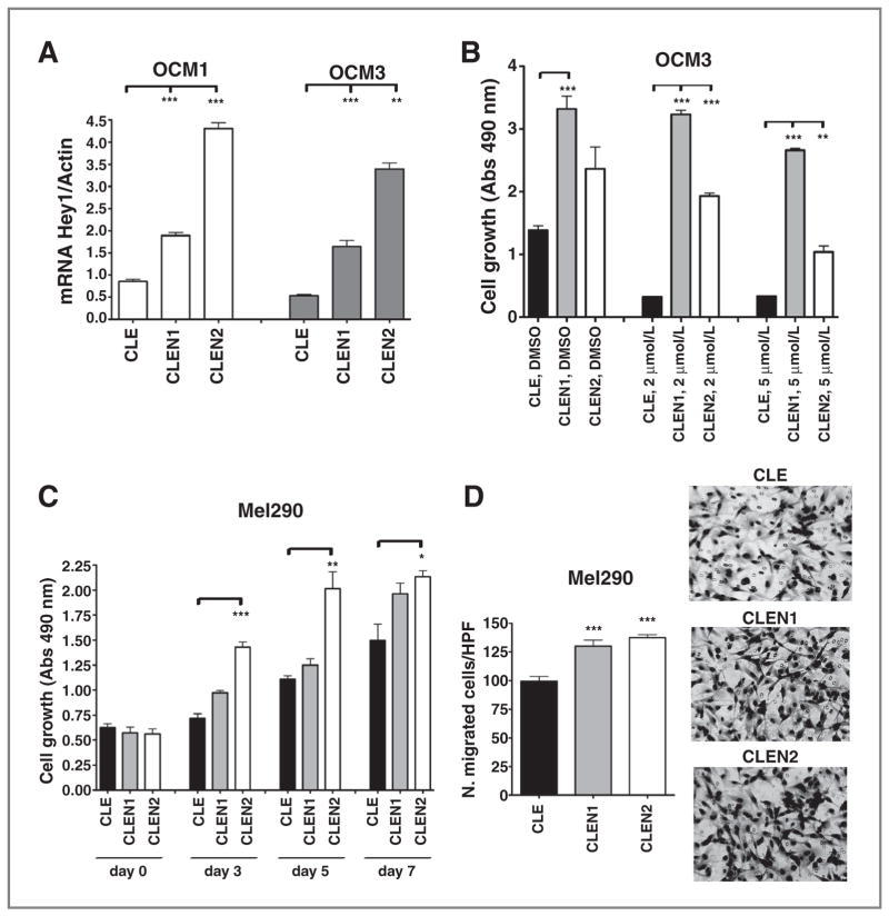
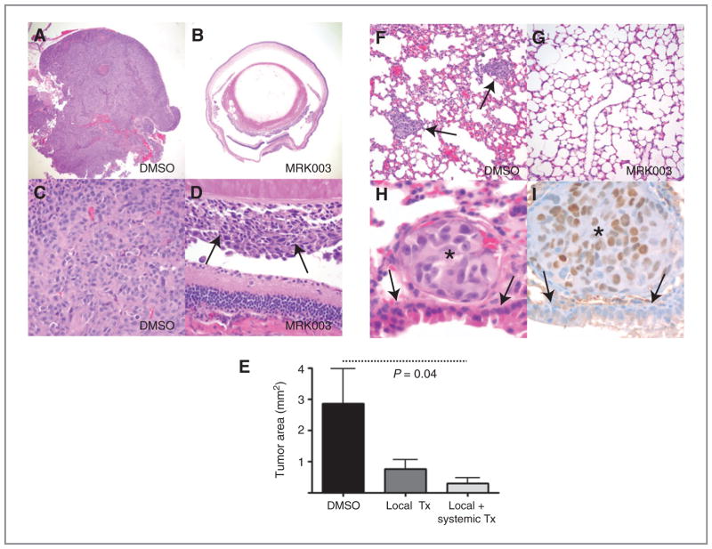
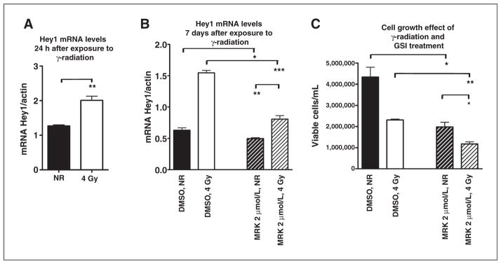
Results: Notch receptors, ligands, and targets were expressed in all five cell lines examined and in 30 primary uveal melanoma samples. Interestingly, the three lines with high levels of baseline pathway activity (OCM1, OCM3, and OCM8) had their growth reduced by pharmacologic Notch blockade using the γ-secretase inhibitor (GSI) MRK003. In contrast, two uveal melanoma lines (Mel285 and Mel290) with very low expression of Notch targets were insensitive to the GSI. Constitutively active forms of Notch1 and Notch2 promoted growth of uveal melanoma cultures and were able to rescue the inhibitory effects of GSI. MRK003 treatment also inhibited anchorage-independent clonogenic growth and cell invasion and reduced phosphorylation levels of STAT3 and extracellular signal-regulated kinase (Erk)1/2. Suppression of canonical Notch activity using short hairpin RNA targeting Notch2 or CBF1 was also able to reduce tumor growth and invasion. Finally, intraocular xenograft growth was significantly decreased by GSI treatment.

Conclusion: Our findings suggest that Notch plays an important role in inducing proliferation and invasion in uveal melanoma and that inhibiting this pathway may be effective in preventing tumor growth and metastasis.


Conflict of interest statement

Disclosure of Potential Conflicts of Interest

C.G. Eberhart discloses a patent licensed to Stemline Therapeutics. No potential conflicts of interest were disclosed by other authors.

Figures

Figure 1
Figure 1
Notch pathway components are expressed in uveal melanoma cell lines and primary tumors. A, mRNA levels of Hes1 were analyzed by quantitative PCR in 30 snap-frozen primary tumors, 5 uveal melanoma cell lines, and normal cutaneous melanocytes (NMEL). B, mRNA transcripts of additional Notch receptors, ligands, and target genes were detected in the 30 primary uveal melanomas (T) and in normal epidermal melanocytes (NMEL; top); Pearson correlation coefficient R was determined between Hes1 and Notch1, Notch2 mRNA levels in the primary uveal melanomas (bottom). C, nuclear Hes1 immunoreactivity was examined in 73 tumors collected on a tissue microarray with strong Hes1 immunostaining shown in the left and a no primary antibody negative control on the right. D, examination of mRNA expression of the Hes1, Hes5, Hey1, Hey2 genes in 5 uveal melanoma cell lines and normal cutaneous melanocytes revealed that three uveal melanoma lines had elevated levels of multiple Notch targets.
Figure 2
Figure 2
Treatment with MRK003 reduces Hes1 mRNA and protein expression and suppresses uveal melanoma growth and invasion. A and B, Hes1 mRNA and protein levels were reduced in OCM1 cells treated with MRK003 at 2, 5, and 10 μmol/L for 48 hours. C, MTS assay of 5 uveal melanoma cell lines treated with MRK003 at 2 and 5 μmol/L for 7 days revealed that growth was only inhibited in cells with high baseline Notch activity. D, clonogenic growth in soft agar assay was significantly reduced by 2 μmol/L MRK003 (*, P = 0.01; **, P = 0.003; ***, P = 0.0005). E and F, transwell assays revealed significantly decreased invasion of OCM3 cells treated for 24 hours with MRK003 as compared with DMSO at the indicated doses (***, P < 0.0004), but no significant changes in Mel290 cells with lower Notch activity. G, cell-cycle analysis of OCM3 cells after 48-hour treatment with MRK003 at 2, 5, and 10 μmol/L, with data from triplicate experiments in right. H, cell-cycle analysis of OCM3 cells 72 hours after infection with shCBF1. I, Western blot analysis reveals increased levels of cleaved caspase-9 in OCM3 cells treated with MRK003 for 96 hours. GAPDH, glyceraldehyde-3-phosphate dehydrogenase.
Figure 2
Figure 2
Treatment with MRK003 reduces Hes1 mRNA and protein expression and suppresses uveal melanoma growth and invasion. A and B, Hes1 mRNA and protein levels were reduced in OCM1 cells treated with MRK003 at 2, 5, and 10 μmol/L for 48 hours. C, MTS assay of 5 uveal melanoma cell lines treated with MRK003 at 2 and 5 μmol/L for 7 days revealed that growth was only inhibited in cells with high baseline Notch activity. D, clonogenic growth in soft agar assay was significantly reduced by 2 μmol/L MRK003 (*, P = 0.01; **, P = 0.003; ***, P = 0.0005). E and F, transwell assays revealed significantly decreased invasion of OCM3 cells treated for 24 hours with MRK003 as compared with DMSO at the indicated doses (***, P < 0.0004), but no significant changes in Mel290 cells with lower Notch activity. G, cell-cycle analysis of OCM3 cells after 48-hour treatment with MRK003 at 2, 5, and 10 μmol/L, with data from triplicate experiments in right. H, cell-cycle analysis of OCM3 cells 72 hours after infection with shCBF1. I, Western blot analysis reveals increased levels of cleaved caspase-9 in OCM3 cells treated with MRK003 for 96 hours. GAPDH, glyceraldehyde-3-phosphate dehydrogenase.
Figure 3
Figure 3
Notch signaling inhibition reduces cell growth and invasion. A and C, MTS assay revealed cell growth inhibition in shNotch2 (A) or shCBF1 (C) infected cells compared with scrambled shRNA or PLKO.1 empty vector infected cells after 5 days of culture (*, P = 0.04; **, P = 0.003; ***, P < 0.0001). B and D, transwell assay revealed that cell invasion was reduced in cells infected with shNotch2 (B) or shCBF1 (D) infected cells compared with scrambled shRNA. P values were calculated versus scrambled shRNA (***, P < 0.0001).
Figure 4
Figure 4
Constitutive Notch activation promotes growth and rescues MRK003 inhibitory effects. A, Hey1 mRNA levels were increased in OCM1 and OCM3 cells after infection with CLEN1 or CLEN2 constructs (**, P = 0.003; ***, P = 0.0005). B, MTS assay conducted in OCM3 cells infected with CLE, CLEN1, or CLEN2 vectors, after 7 days of treatment with MRK003, shows that growth inhibition induced by GSI is rescued by Notch pathway activation (**, P = 0.002; ***, P < 0.0008). C and D, CLEN1 and CLEN2 constructs also promote cell growth and invasion as determined respectively by MTS (C) and Transwell (D) assays in Mel290 cells (*, P = 0.02; **, P < 0.006; ***, P = 0.0009).
Figure 5
Figure 5
Notch pathway inhibition affects lung metastasis. A and B, a large tumor filled the entire eye 3 weeks after intravitreal injection of OCM1 cells treated with vehicle (DMSO), whereas only microscopic disease was present in most eyes receiving MRK003 at the time of xenograft initiation (original magnification 40× for both panels). C and D, higher magnification views showing solid sheets of melanoma cells in control eyes, but only focal tumor growth (D, arrows) following local MRK003 treatment (original magnification 400×). E, intraocular xenograft area was significantly reduced (P = 0.04) by combined local and systemic MRK003 treatment, whereas the changes in tumors treated locally alone was not significant. F and G, multiple melanoma metastases were identified in the lungs of 2 of 10 control animals (F, arrows), but none in the 20 mice treated with MRK003 (G, original magnification 200×). H and I, the metastatic tumor nodules were composed of cells with enlarged nuclei and prominent nucleoli (asterisks) as compared with normal adjacent bronchial cells (arrows, original magnification 1,000×). Immunostains for the MITF were positive in tumor cells (I).
Figure 6
Figure 6
Combination of MRK003 treatment with γ-radiation. A, Hey1 mRNA levels were significantly increased in OCM1 cells 24 hours after exposure to 4 Gy of γ-radiation as compared with nonirradiated cells (NR) cells (**, P < 0.005). B, Hey1 mRNA levels remained elevated 7 days after radiation, but could be suppressed by MRK003 (*, P = 003; **, P = 0.006; ***, P = 0.0004). C, OCM1 cells were treated with 2 μmol/L MRK003 or DMSO for 5 hours and then exposed to 0 or 4 Gy of γ-radiation in a single dose. Viable cells were counted 7 days after radiation exposure in triplicate experiments (*, P = 0.045; **, P = 0.01).

References

    1. Landreville S, Agapova OA, Harbour JW. Emerging insights into the molecular pathogenesis of uveal melanoma. Future Oncol. 2008;4:629–36. - PMC - PubMed
    1. Shields CL, Furuta M, Thangappan A, Nagori S, Mashayekhi A, Lally DR, et al. Metastasis of uveal melanoma millimeter-by-millimeter in 8033 consecutive eyes. Arch Ophthalmol. 2009;127:989–98. - PubMed
    1. Prescher G, Bornfeld N, Hirche H, Horsthemke B, Jockel KH, Becher R. Prognostic implications of monosomy 3 in uveal melanoma. Lancet. 1996;347:1222–5. - PubMed
    1. Damato B, Dopierala JA, Coupland SE. Genotypic profiling of 452 choroidal melanomas with multiplex ligation-dependent probe amplification. Clin Cancer Res. 2010;16:6083–92. - PubMed
    1. Shields CL, Ganguly A, Bianciotto CG, Turaka K, Tavallali A, Shields JA. Prognosis of uveal melanoma in 500 cases using genetic testing of fine-needle aspiration biopsy specimens. Ophthalmology. 2011;118:396–401. - PubMed

Publication types

MeSH terms