. 2012 Mar;30(3):405-14.
doi: 10.1002/stem.1020.

REST regulates oncogenic properties of glioblastoma stem cells

Affiliations

REST regulates oncogenic properties of glioblastoma stem cells

Mohamed M Kamal et al. Stem Cells. 2012 Mar.

Erratum in

  • Stem Cells. 2012 May;30(5):1049

Abstract

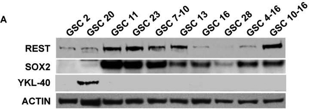
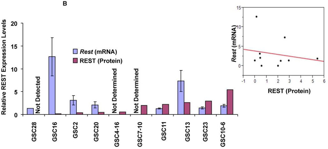
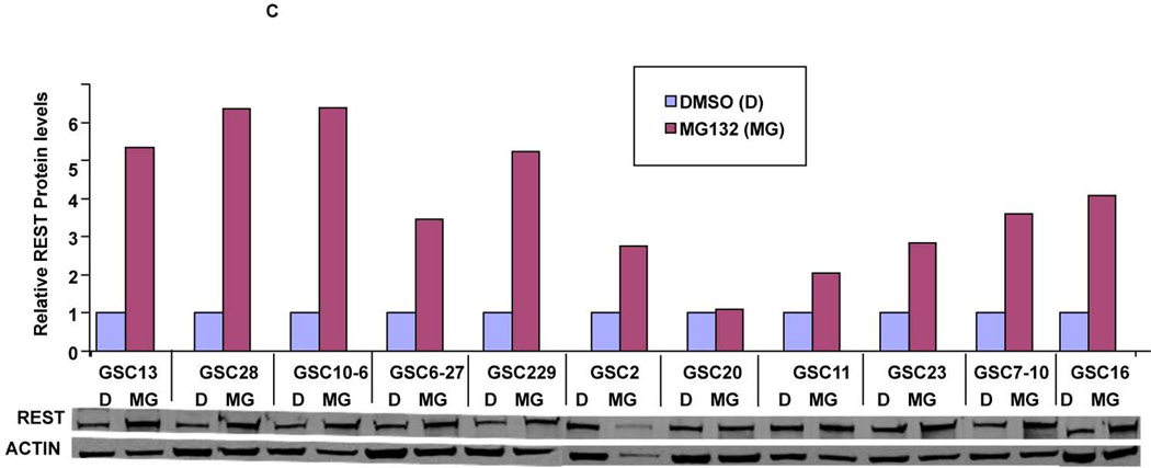
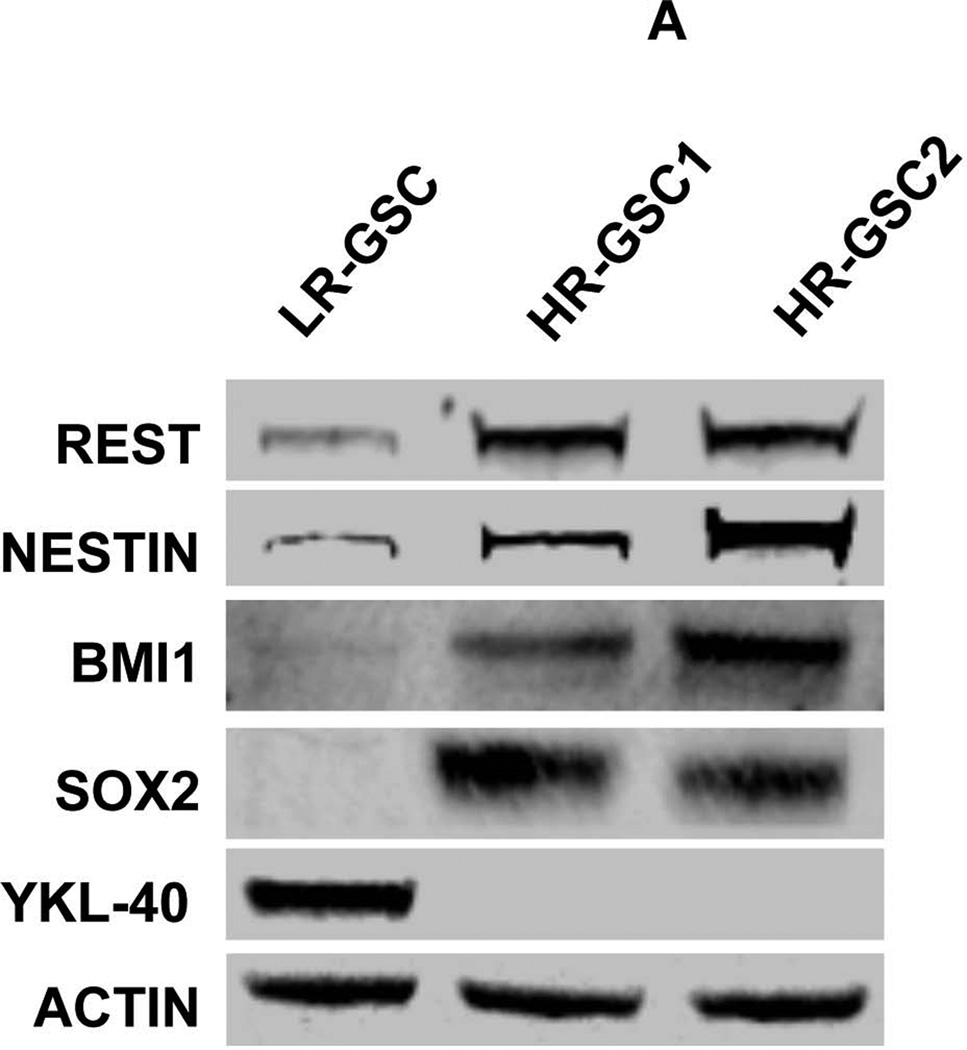
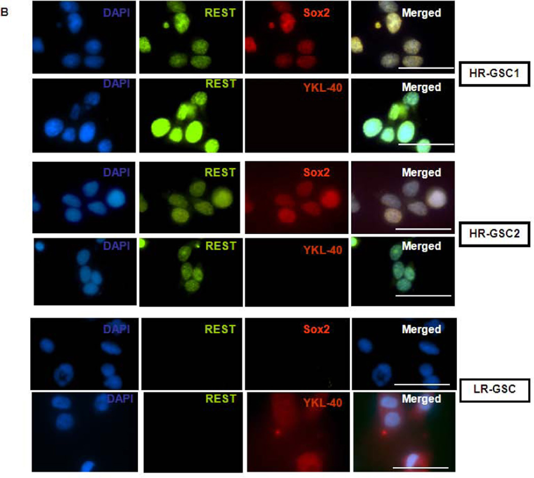
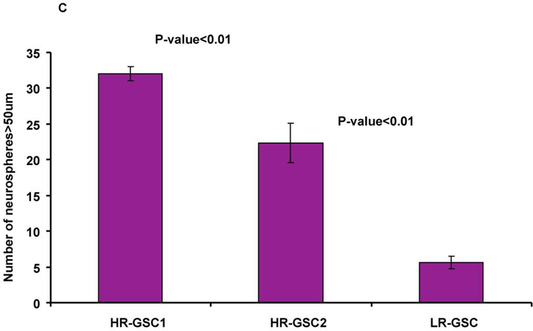
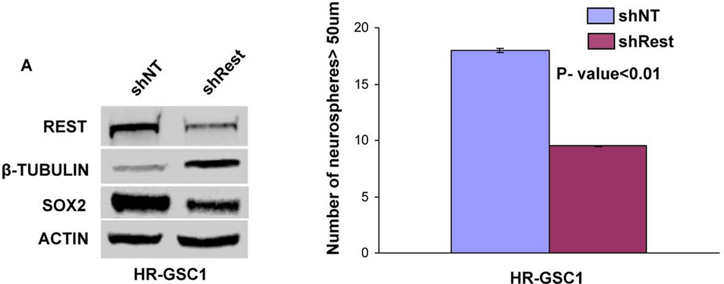
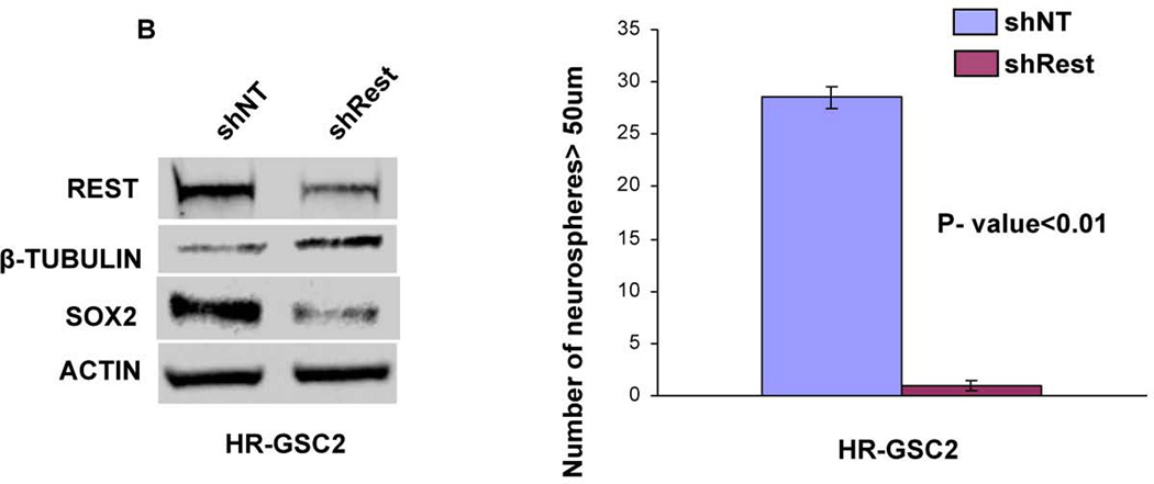
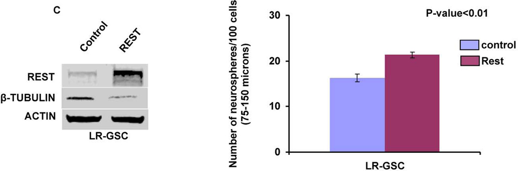
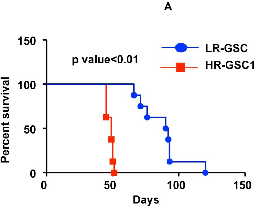
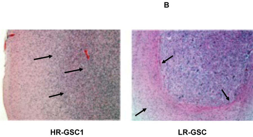
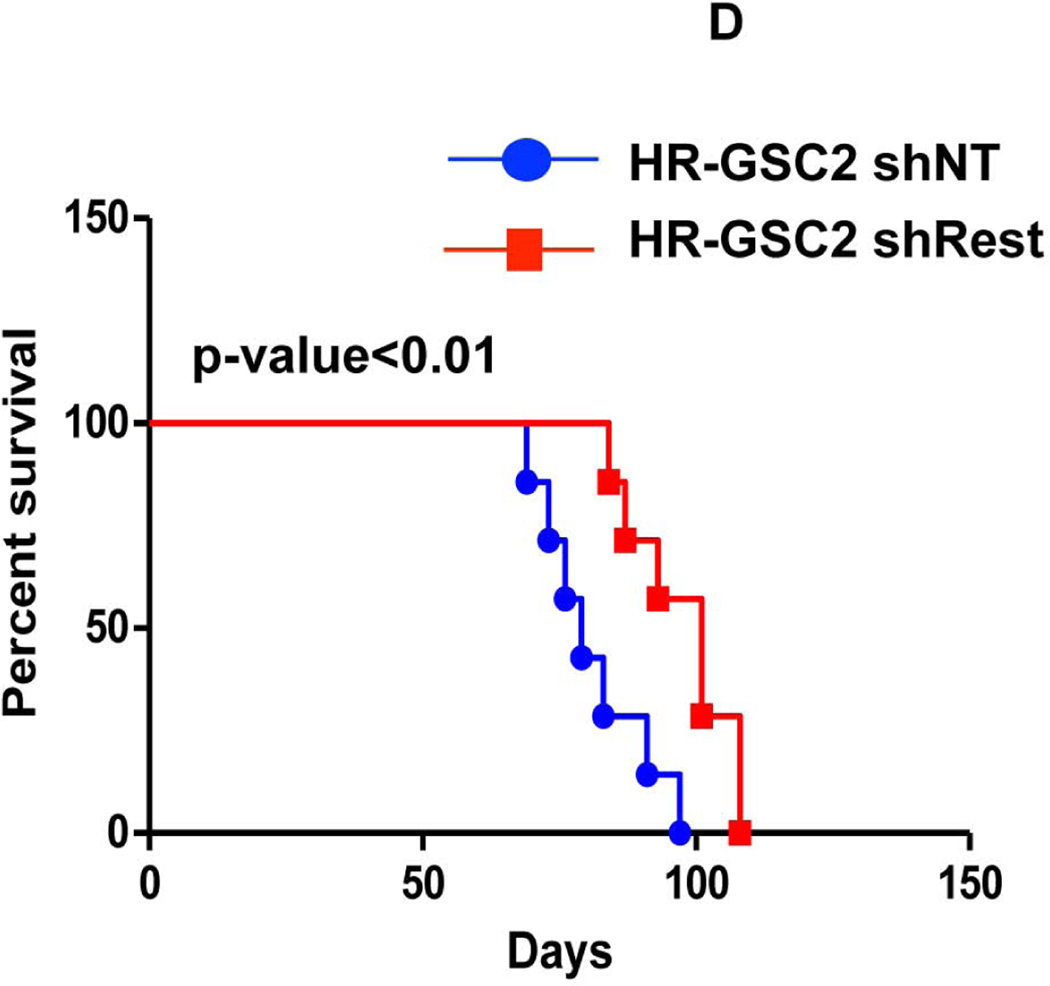
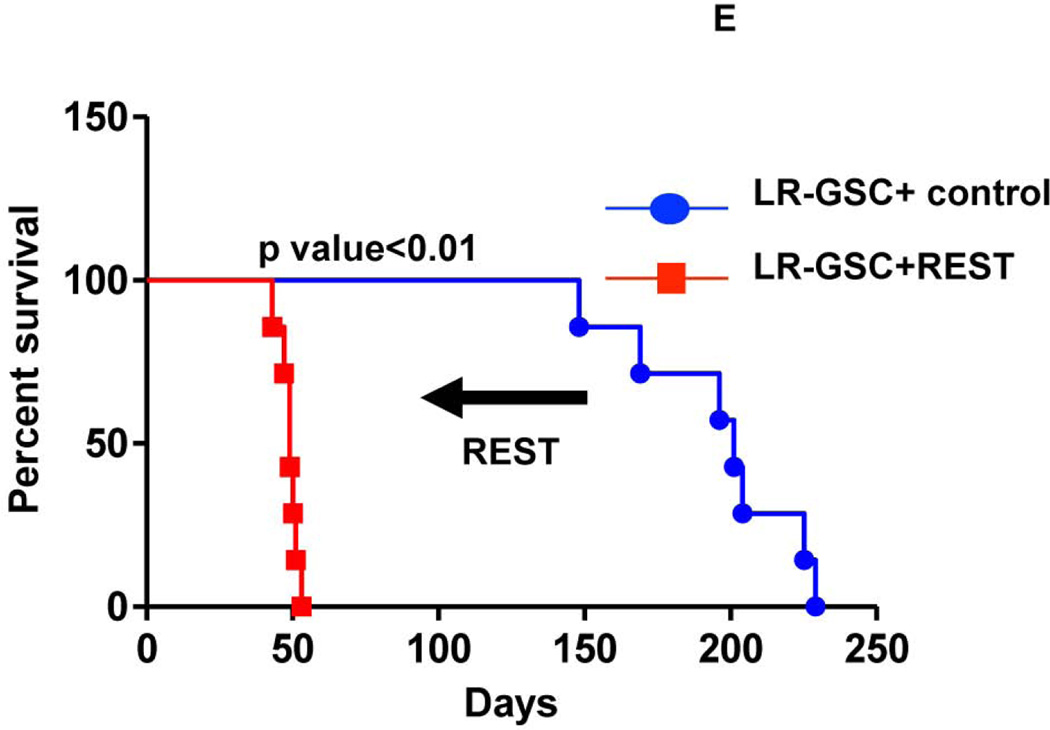
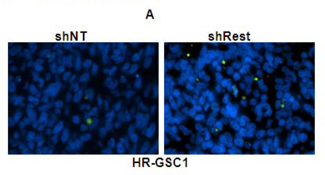
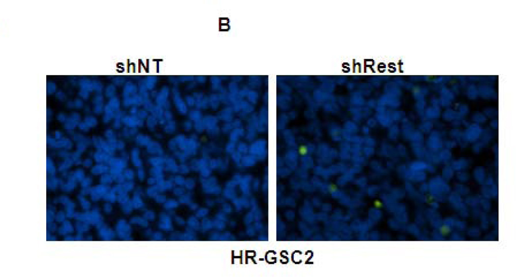
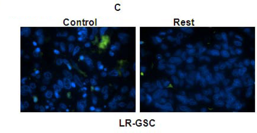
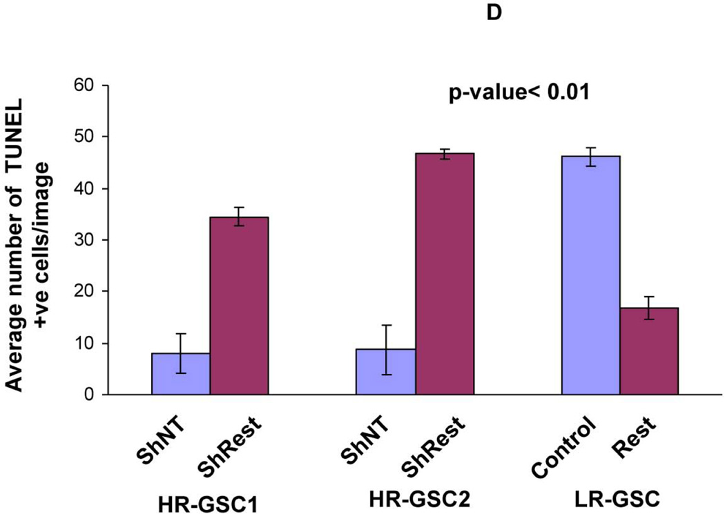
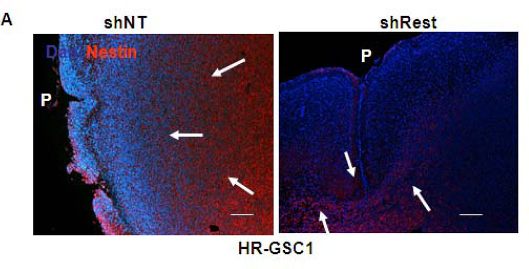
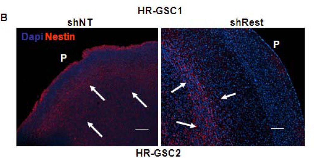
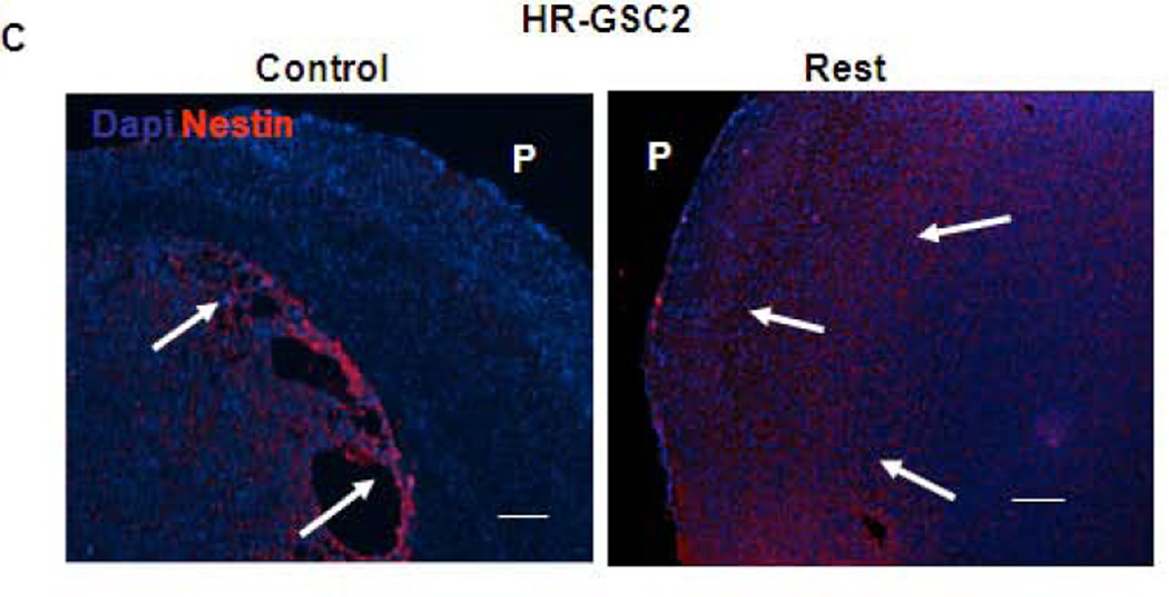
Glioblastoma multiforme (GBM) tumors are the most common malignant primary brain tumors in adults. Although many GBM tumors are believed to be caused by self-renewing, glioblastoma-derived stem-like cells (GSCs), the mechanisms that regulate self-renewal and other oncogenic properties of GSCs are only now being unraveled. Here we showed that GSCs derived from GBM patient specimens express varying levels of the transcriptional repressor repressor element 1 silencing transcription factor (REST), suggesting heterogeneity across different GSC lines. Loss- and gain-of-function experiments indicated that REST maintains self-renewal of GSCs. High REST-expressing GSCs (HR-GSCs) produced tumors histopathologically distinct from those generated by low REST-expressing GSCs (LR-GSCs) in orthotopic mouse brain tumor models. Knockdown of REST in HR-GSCs resulted in increased survival in GSC-transplanted mice and produced tumors with higher apoptotic and lower invasive properties. Conversely, forced expression of exogenous REST in LR-GSCs produced decreased survival in mice and produced tumors with lower apoptotic and higher invasive properties, similar to HR-GSCs. Thus, based on our results, we propose that a novel function of REST is to maintain self-renewal and other oncogenic properties of GSCs and that REST can play a major role in mediating tumorigenicity in GBM.


Figures

Figure 1
Figure 1. (A) Patient glioblastoma-derived stem cells (GSCs) show varying REST levels
Western blotting assays of GSCs obtained from 10 human GBM patient specimens showed varying levels of REST, Sox2 and YKL-40 proteins. Actin was used as an internal control. (B) Comparison of REST expression at the protein and transcript levels indicated that REST protein levels do not correspond to Rest transcript levels (left panel). A scatter plot is also shown (Right panel, Rsq=0.04). (C) Western blot based quantification of REST protein levels in GSCs treated with either the proteasomal inhibitor (MG132) or solvent (DMSO) indicated that REST protein is sensitive to proteasomal degradation in most of the GSC lines.
Figure 2
Figure 2. High REST protein expression correlates with stemness markers in GSCs
(A) Expression of REST, Nestin, Bmi1, Sox2, and YKL40 in two HR-GSCs (HR-GSC1 and HR-GSC2) and one LR-GSC were determined by Western blotting assays. Actin was used as an internal control. (B) Immunofluorescence analysis of the two HR-GSCs and one LR-GSC shows a distinct pattern of expression of Sox2, and YKL-40. The bar in the figures represents 50 micron. (C) HR-GSCs (HR-GSC1 and HR-GSC2) show higher neurosphere-formation ability compared to LR-GSCs.
Figure 3
Figure 3. REST controls self-renewal of GSCs
(A, B) Rest loss-of-function experiments. Knockdown of REST with shRest (shNT as a control) in HR-GSC1 (A) and HR-GSC2 (B) caused lowered expression of REST, higher expression of the direct REST target neuronal beta-tubulin and lower expression of neural stem cell self-renewal regulator Sox2 as determined by Western blotting assay (left panels), and lowered self-renewal ability (right panels) (n=48). (C) REST gain-of-function experiments. Expression of exogenous REST in LR-GSCs (expression of exogenous GFP served as a control) caused decreased β-tubulin expression (left panel) and increased self-renewal ability (right panel) (n=48).
Figure 4
Figure 4. REST regulates survival in mice bearing GSC-mediated brain tumors
(A, B) HR-GSC1 and LR-GSC cells were transplanted into the brains of nude mice, the mice were assayed for survival using Kaplan-Meier method (A) and histopathology of the resulting tumors were examined by H&E staining (B) (n=7 mice per treatment group). HR-GSC1 tumors produced shorter survival in mice than LR-GSC tumors and HR-GSC1 tumors were more infiltrative than LR-GSC tumors, which showed circumscribed masses. Arrows in the same direction indicate more infiltrative cells while those in the opposite direction represent more restrictive structure. (C, D) Rest loss-of-function tumors. Stable lines of HR-GSC1 (C) and HR-GSC2 (D) cells expressing either shREST or shNT were transplanted into the brains of nude mice and mouse survival was compared. Knockdown of REST produced increased survival in tumor-bearing mice generated by both lines (n=7 mice per treatment group). (D) Rest gain-of-function tumors. LR-GSCs expressing either exogenous REST or GFP were transplanted into the brains of nude mice, the mice were assayed for survival. Additional REST converted LR-GSCs into more infiltrative tumors and decreased survival in tumor-bearing mice (n=7 mice per treatment group).
Figure 5
Figure 5. REST suppresses apoptosis in GSC-mediated brain tumors
(A–C) In vivo TUNEL assay indicating increased apoptosis in mouse tumors upon knockdown of REST in HR-GSC1 (A) and HR-GSC2 (B). LR-GSCs showed reduced apoptosis upon expression of exogenous REST compared to exogenous GFP control (C). (D) quantification of in vivo TUNEL assays shown in A–C.
Figure 6
Figure 6. REST regulates invasion in GSC-mediated brain tumors
(A–C) Both HR-GSC1 (A)- and HR-GSC2 (B)-mediated tumors showed decreased invasion upon shRest-treatment compared to control shNT-treatment. LR-GSC-mediated tumors showed increased invasion upon expression of exogenous REST compared to GFP control (C). Arrows in the same direction indicate more infiltrative cells while those in the opposite direction represent more restrictive structure.P = pial layer. Bar = 50 micron.

Similar articles

Cited by

References

    1. Furnari FB, Fenton T, Bachoo RM, et al. Malignant astrocytic glioma: genetics, biology, and paths to treatment. Genes Dev. 2007;21:2683–2710. - PubMed
    1. Gladson CL, Prayson RA, Liu WM. The pathobiology of glioma tumors. Annu Rev Pathol. 2010;5:33–50. - PMC - PubMed
    1. Louis DN, Ohgaki H, Wiestler OD, et al. The 2007 WHO classification of tumours of the central nervous system. Acta Neuropathol. 2007;114:97–109. - PMC - PubMed
    1. Stupp R, Hegi ME, Mason WP, et al. Effects of radiotherapy with concomitant and adjuvant temozolomide versus radiotherapy alone on survival in glioblastoma in a randomised phase III study: 5-year analysis of the EORTC-NCIC trial. The Lancet Oncology. 2009;10:459–466. - PubMed
    1. Gilbertson RJ, Gutmann DH. Tumorigenesis in the brain: location, location, location. Cancer Res. 2007;67:5579–5582. - PubMed

Publication types

MeSH terms