Comparative Study
. 2012 Mar;194(6):1299-306.
doi: 10.1128/JB.06474-11. Epub 2012 Jan 6.

Three-dimensional structures of pathogenic and saprophytic Leptospira species revealed by cryo-electron tomography

Affiliations
Comparative Study

Three-dimensional structures of pathogenic and saprophytic Leptospira species revealed by cryo-electron tomography

Gianmarco Raddi et al. J Bacteriol. 2012 Mar.

Abstract

Leptospira interrogans is the primary causative agent of the most widespread zoonotic disease, leptospirosis. An in-depth structural characterization of L. interrogans is needed to understand its biology and pathogenesis. In this study, cryo-electron tomography (cryo-ET) was used to compare pathogenic and saprophytic species and examine the unique morphological features of this group of bacteria. Specifically, our study revealed a structural difference between the cell envelopes of L. interrogans and Leptospira biflexa involving variations in the lipopolysaccharide (LPS) layer. Through cryo-ET and subvolume averaging, we determined the first three-dimensional (3-D) structure of the flagellar motor of leptospira, with novel features in the flagellar C ring, export apparatus, and stator. Together with direct visualization of chemoreceptor arrays, DNA packing, periplasmic filaments, spherical cytoplasmic bodies, and a unique "cap" at the cell end, this report provides structural insights into these fascinating Leptospira species.


Figures

Fig 1
Fig 1
Ultrastructure from 3-D reconstruction of intact L. interrogans (A) or L. biflexa (B). The outer membrane (OM), inner membrane (IM), peptidoglycan layer (PG), and periplasmic flagellum (PF), the “cap” at the cell end, and a spherical body (SB) can be discerned from the picture. Zoom-in views reveal the structural details of the cell envelope of L. interrogans (C) or L. biflexa (D), respectively. LPS extends from the outer leaflet to a strong continuous density layer. Density profiles across the membranes of L. interrogans and L. biflexa (dashed line) are shown in panels E and F, respectively. The LPS layer was considerably thicker in L. interrogans than that in L. biflexa.
Fig 2
Fig 2
A gallery of the cell end reconstructions reveals a “cap”-like feature of L. biflexa (A to D) and L. interrogans (E to H).
Fig 3
Fig 3
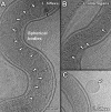
Periplasmic filaments in Leptospira spp. (A) Periplasmic filaments (PFil) are visible in the middle of the elongated organisms, while PF of Leptospira is located near the termini. (B) An enlarged section from panel A of PFil in L. interrogans. Three filaments are observed within the boundary of the outer membrane (OM). (C) A cross-sectional view indicates that PFil are located between the outer membrane (OM) and the inner membrane (IM).
Fig 4
Fig 4
Molecular architecture of the intact flagellar motor of Leptospira spp. (A) Centered section parallel to the direction of the filament of an asymmetric reconstruction of the Leptospiral motors. Panels B, C, D, and E are horizontal cross sections. The locations of the sections are indicated in panel A. Panel B shows a section that lies inside the socket formed by the collar in periplasmic space. Section C transects the cytoplasmic side of the MS ring, just above the C ring. Sections D and E are located on the top and bottom of the C ring, respectively. Surface rendering of the Leptospira flagellar motor is presented in panels F and G. The side view (F and G) shows a novel C-ring structure. Extra density (in pink) is found at the bottom of the C ring. In panel G, 16 ring-like structures (colored in blue) are located on top of the C ring. Each ring is about 7.3 nm in diameter and 7.5 nm in height. The top portion of the ring is embedded in the cytoplasmic membrane, while the bottom portion is likely interacting with the C ring. Panel H is a view 45° rotated from panel G, revealing the interaction between the C ring and the stators.
Fig 5
Fig 5
Visualization of chemoreceptor arrays of Leptospira spp. Putative chemoreceptor arrays were observed in L. interrogans (A) and L. biflexa (B). The insets are the corresponding zoom-in views of the arrays outlined with white dashed lines. The arrays appear as clusters of pillar-like densities that extend from the IM and connect with a layer of high electron density at the membrane-distal ends. These are likely formed by CheA/W, which is known to form a continuous layer at the bottom of the chemoreceptor arrays. An additional array, unique to the saprophytic species, is found at the cell end (C). The inset is the zoom-in image of the array, indicating the presence of extra density layers (EL). The distance between the IM and the basal layer of CheA/W is relatively constant (25 nm) for most of the arrays, as shown in the cross sections (D and E). However, the novel array is longer (33 nm), as shown in the cross sections (F). The scale bar is 50 nm.
Fig 6
Fig 6
Two types of spherical bodies in Leptospira spp. High- and low-density spherical bodies were labeled with dark and white arrows, respectively. (A) Cryo-ET tomogram slice of an L. biflexa cell, with spherical bodies (∼100 nm in size) located in the cell center. (B) Cryo-ET of an L. interrogans cell shows that the spherical bodies are randomly positioned along the cell body, with a diameter of ∼30 nm. (C) High-magnification view of a free-in-solution spherical body showing the absence of a phospholipid bilayer.
Fig 7
Fig 7
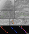
Putative DNA bundles in intact and lysed cells and in cell medium. (A) Cryo-ET of L. biflexa cell reveals putative DNA bundles in the cell body. (B) Image of a partially lysed L. interrogans cell, showing that the putative DNA maintains a filamentous structure. Panels C and D show the zoom-in views of the areas highlighted in panels A and B, respectively. The contour lines outline the boundary along the putative DNA structure. (E) Top view of the putative DNA bundle in cell medium. (F) Zoom-in view of area indicated in panel E. Orange arrows depict putative DNA strands. (G) Cross-section view of the putative DNA bundle shown in panel F. The bundle is tightly packed by filaments with a spacing of ∼4 nm and a diameter of ∼2 nm. Fluorescence microscopy shows continuous dyed DNA in blue (I), cell envelope in red (H), and a composite image (J).

Similar articles

Cited by

References

    1. Beck M, et al. 2009. Visual proteomics of the human pathogen Leptospira interrogans. Nat. Methods 6:817–823 - PMC - PubMed
    1. Bharti AR, et al. 2003. Leptospirosis: a zoonotic disease of global importance. Lancet Infect. Dis. 3:757–771 - PubMed
    1. Bloomfield VA. 1996. DNA condensation. Curr. Opin. Struct. Biol. 6:334–341 - PubMed
    1. Briegel A, et al. 2009. Universal architecture of bacterial chemoreceptor arrays. Proc. Natl. Acad. Sci. U. S. A. 106:17181–17186 - PMC - PubMed
    1. Bromley DB, Charon NW. 1979. Axial filament involvement in the motility of Leptospira interrogans. J. Bacteriol. 137:1406–1412 - PMC - PubMed

Publication types

MeSH terms

Substances