. 2012 Feb 24;287(9):6318-25.
doi: 10.1074/jbc.M111.331090. Epub 2012 Jan 6.

Mechanism for release of alkaline phosphatase caused by glycosylphosphatidylinositol deficiency in patients with hyperphosphatasia mental retardation syndrome

Affiliations

Mechanism for release of alkaline phosphatase caused by glycosylphosphatidylinositol deficiency in patients with hyperphosphatasia mental retardation syndrome

Yoshiko Murakami et al. J Biol Chem. .

Abstract

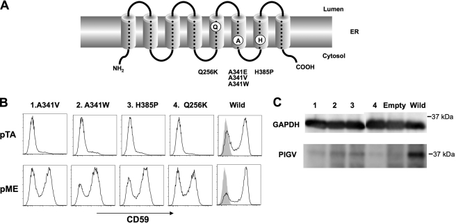
Hyperphosphatasia mental retardation syndrome (HPMR), an autosomal recessive disease characterized by mental retardation and elevated serum alkaline phosphatase (ALP) levels, is caused by mutations in the coding region of the phosphatidylinositol glycan anchor biosynthesis, class V (PIGV) gene, the product of which is a mannosyltransferase essential for glycosylphosphatidylinositol (GPI) biosynthesis. Mutations found in four families caused amino acid substitutions A341E, A341V, Q256K, and H385P, which drastically decreased expression of the PIGV protein. Hyperphosphatasia resulted from secretion of ALP, a GPI-anchored protein normally expressed on the cell surface, into serum due to PIGV deficiency. In contrast, a previously reported PIGM deficiency, in which there is a defect in the transfer of the first mannose, does not result in hyperphosphatasia. To provide insights into the mechanism of ALP secretion in HPMR patients, we took advantage of CHO cell mutants that are defective in various steps of GPI biosynthesis. Secretion of ALP requires GPI transamidase, which in normal cells, cleaves the C-terminal GPI attachment signal peptide and replaces it with GPI. The GPI-anchored protein was secreted substantially into medium from PIGV-, PIGB-, and PIGF-deficient CHO cells, in which incomplete GPI bearing mannose was accumulated. In contrast, ALP was degraded in PIGL-, DPM2-, or PIGX-deficient CHO cells, in which incomplete shorter GPIs that lacked mannose were accumulated. Our results suggest that GPI transamidase recognizes incomplete GPI bearing mannose and cleaves a hydrophobic signal peptide, resulting in secretion of soluble ALP. These results explain the molecular mechanism of hyperphosphatasia in HPMR.


Figures

FIGURE 1.
FIGURE 1.
A, PIGV mutations found in patients with HPMR syndrome. B, inefficient restoration of GPI-AP expression on PIGV-deficient CHO cells by mutant PIGV cDNA. PIGV-deficient CHO cells were transiently transfected with weak promoter-driven pTA PIGV constructs (upper panel) or strong promoter-driven pME PIGV constructs (lower panel) carrying indicated mutations or a wild-type construct or an empty vector (gray shadow) and were analyzed for the surface expression of CD59 by FACS. C, decreased expression of mutant PIGV. Expressions of FLAG-tagged PIGV protein were analyzed by Western blotting 2 days after transfection with strong promoter-driven pME PIGV constructs. Number of each lane indicates each mutant shown in B; GAPDH is a loading control.
FIGURE 2.
FIGURE 2.
A, surface expression of PLAP after cotransfection with PIGV into PIGV-deficient CHO cells. PIGV-deficient CHO cells were transiently transfected with pME HA-PLAP together with pME PIGV (dotted line) or empty vector (solid line) and analyzed for the surface expression of PLAP by FACS. Shadow, without PLAP expression vector. B, ALP activity in culture medium and cell lysate after cotransfection of PLAP and PIGV cDNAs into PIGV-deficient CHO cells. Each transfectant in A was analyzed. Relative ALP activity in culture medium (black bars), cell lysates (dark gray bars), and total (light gray bars) against the total ALP activity in PIGV-rescued CHO cell. Each value indicates the mean ± S.D. of four independent experiments.
FIGURE 3.
FIGURE 3.
Pulse-chase metabolic labeling of GPI-APs. A, HA-PLAP expressing PIGV-deficient cells (left panel), PIGV-rescued (middle panel) and PIGK-deficient (right panel) CHO cells were pulsed with [35S]methionine and [35S]cysteine for 10 min and then chased for the indicated periods. The cell lysates and culture supernatants were immunoprecipitated with an anti-HA antibody, separated by SDS-PAGE, and analyzed using a BAS 1000 analyzer. B, DAF expressing PIGV-deficient cells (left panel), PIGV-rescued (middle panel), and PIGK-deficient (right panel) CHO cells were analyzed as in A. C, quantified data of B. Intensity of total labeled protein at the start of the chase was 100%. Broken lines, ER form; solid lines, secreted DAF; dotted lines, mature form.
FIGURE 4.
FIGURE 4.
A, identification and characterization of C-terminal peptide by LC/ESI MS/MS. Top, spectra of peptides eluted from LC. The peak at the retention time of 21.72 min contained a C-terminal peptide cleaved at the ω site in the GPI attachment signal; bottom left, the first MS analysis of the peptides eluted at 21.72 min; bottom right, MS/MS spectrum of a parent ion with m/z 545.252+ and its sequence. The b-series fragments are indicated in bold. B, the ω site-dependent release of PLAP from PIGV-deficient CHO cells. PIGV-deficient CHO cells were transiently cotransfected with wild-type (dark gray bar) or ω site mutant (light gray bar) HA-PLAP and luciferase reporter construct for normalization. Relative PLAP activities in the media are shown.
FIGURE 5.
FIGURE 5.
Secretion of PLAP from CHO cell mutants defective in various GPI biosynthesis steps. A, various CHO cell mutants were transiently transfected with PLAP expression vector and the luciferase reporter construct for normalization together with cDNA of each responsible gene or empty vector. Normalized relative PLAP activities in the medium from various mutants are shown. B, GPI biosynthesis pathway. Genes defective in CHO cell mutants used in A are shown in bold. Modified from Ref. .
FIGURE 6.
FIGURE 6.
Models of different processing of GPI-AP proprotein in wild-type and GPI-defective cells. In wild-type cells, the GPI transamidase complex recognizes both the GPI attachment signal and GPI, and processes the proprotein at the ω site and attaches it to GPI. GPI-APs are expressed on the cell surface and a small fraction of GPI-AP is released by some GPI-cleaving enzyme (top). In late step mutants, a significant fraction of proprotein is processed at the ω site by the GPI transamidase, and is released into the culture medium, whereas the rest of the proprotein is degraded (second top). In early step mutants, GPI transamidase is not activated; bound proprotein is mostly degraded and a small fraction is released by an unknown mechanism (second bottom). In GPI transamidase mutants, proprotein is completely degraded within the cell (bottom).

References

    1. Krawitz P. M., Schweiger M. R., Rödelsperger C., Marcelis C., Kölsch U., Meisel C., Stephani F., Kinoshita T., Murakami Y., Bauer S., Isau M., Fischer A., Dahl A., Kerick M., Hecht J., Köhler S., Jäger M., Grünhagen J., de Condor B. J., Doelken S., Brunner H. G., Meinecke P., Passarge E., Thompson M. D., Cole D. E., Horn D., Roscioli T., Mundlos S., Robinson P. N. (2010) Identity-by-descent filtering of exome sequence data identifies PIGV mutations in hyperphosphatasia mental retardation syndrome. Nat. Genet. 42, 827–829 - PubMed
    1. Kang J. Y., Hong Y., Ashida H., Shishioh N., Murakami Y., Morita Y. S., Maeda Y., Kinoshita T. (2005) PIG-V involved in transferring the second mannose in glycosylphosphatidylinositol. J. Biol. Chem. 280, 9489–9497 - PubMed
    1. Thompson M. D., Nezarati M. M., Gillessen-Kaesbach G., Meinecke P., Mendoza-Londono R., Mornet E., Brun-Heath I., Squarcioni C. P., Legeai-Mallet L., Munnich A., Cole D. E. (2010) Hyperplasia with seizures, neurologic deficit, and characteristic facial features: Five new patients with Mabry syndrome. Am. J. Med. Genet. A 152, 1661–1669 - PubMed
    1. Ohishi K., Inoue N., Kinoshita T. (2001) PIG-S and PIG-T, essential for GPI anchor attachment to proteins, form a complex with GAA1 and GPI8. EMBO J. 20, 4088–4098 - PMC - PubMed
    1. Hong Y., Ohishi K., Kang J. Y., Tanaka S., Inoue N., Nishimura J., Maeda Y., Kinoshita T. (2003) Human PIG-U and yeast Cdc91p are the fifth subunit of GPI transamidase that attaches GPI anchors to proteins. Mol. Biol. Cell 14, 1780–1789 - PMC - PubMed

Publication types

MeSH terms

Supplementary concepts