. 2012 Mar 2;287(10):7367-73.
doi: 10.1074/jbc.M111.316828. Epub 2012 Jan 6.

Reductive activation of type 2 ribosome-inactivating proteins is promoted by transmembrane thioredoxin-related protein

Affiliations

Reductive activation of type 2 ribosome-inactivating proteins is promoted by transmembrane thioredoxin-related protein

Matteo Pasetto et al. J Biol Chem. .

Abstract

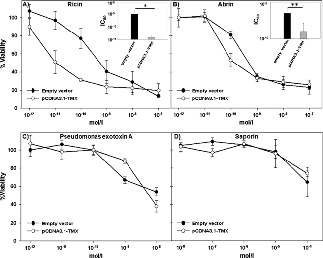
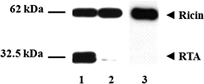
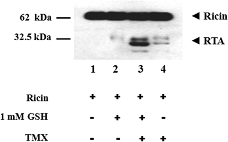
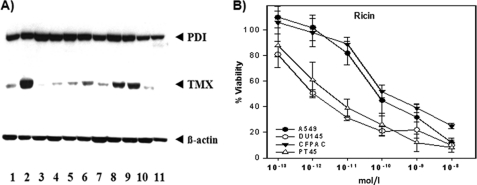
Members of the type 2 ribosome-inactivating proteins (RIPs) family (e.g. ricin, abrin) are potent cytotoxins showing a strong lethal activity toward eukaryotic cells. Type 2 RIPs contain two polypeptide chains (usually named A, for "activity", and B, for "binding") linked by a disulfide bond. The intoxication of the cell is a consequence of a reductive process in which the toxic domain is cleaved from the binding domain by oxidoreductases located in the lumen of the endoplasmic reticulum (ER). The best known example of type 2 RIPs is ricin. Protein disulfide isomerase (PDI) was demonstrated to be involved in the process of ricin reduction; however, when PDI is depleted from cell fraction preparations ricin reduction can still take place, indicating that also other oxidoreductases might be implicated in this process. We have investigated the role of TMX, a transmembrane thioredoxin-related protein member of the PDI family, in the cell intoxication operated by type 2 RIPs ricin and abrin. Overexpressing TMX in A549 cells resulted in a dramatic increase of ricin or abrin cytotoxicity compared with control mock-treated cells. Conversely, no difference in cytotoxicity was observed after treatment of A549 cells or control cells with saporin or Pseudomonas exotoxin A whose intracellular mechanism of activation is not dependent upon reduction (saporin) or only partially dependent upon it (Pseudomonas exotoxin A). Moreover, the silencing of TMX in the prostatic cell line DU145 reduced the sensitivity of the cells to ricin intoxication further confirming a role for this enzyme in intracellular ricin activation.


Figures

FIGURE 1.
FIGURE 1.
Sensitivity of A549 transfectants (empty vector and pCDNA3.1-TMX) to toxins. A549 control cells (empty vector) and overexpressing TMX (pCDNA3.1-TMX) were exposed to various concentrations of toxins and their viability assessed 48 h after toxin addition by an XTT assay. The graphs represent, respectively, the intoxication of the target cells after incubation with ricin (A), abrin (B), Pseudomonas exotoxin A (C), and saporin (D). In the upper right corner of A and B are reported the histograms of the IC50 (concentration inhibiting the 50% of the cell viability) value obtained from the cytotoxicity curves after treatment of the cells with ricin or abrin. *, p < 0.01; **, <0.05 (Student's t test).
FIGURE 2.
FIGURE 2.
Reduction of ricin with prereduced TMX. Samples with ricin (2 μg) were incubated with 5 μg of DTT-preactivated TMX for 60 min at 37 °C (lane 1) in a final volume of 50 μl. Ricin incubated with TMX without DTT preactivation is represented in lane 2. Ricin alone (lane 3) was included as a control. After 60 min the reaction was stopped by adding loading buffer and running the SDS-PAGE under nonreducing conditions. The appearance of reduced ricin was determined by Western blotting as described under “Results and Discussion.”
FIGURE 3.
FIGURE 3.
Glutathione can activate TMX in vitro. Samples with ricin (2 μg) were incubated for 60 min at 37 °C in the absence (lanes 1 and 2) or in the presence (lanes 3 and 4) of 5 μg of oxidized TMX (final volume of 50 μl). GSH (1 mm) was present in samples of lanes 2 and 3. Samples with ricin alone (lane 1) or without TMX only (lane 2) were included as controls. The reduction of ricin was evaluated by Western blotting as described.
FIGURE 4.
FIGURE 4.
Reduction of ricin in an oxidizing environment. To check the ability of TMX to reduce ricin in different redox conditions we included in the buffer (0.1 m phosphate buffer) different GSSG:GSH ratios. To set up the oxidizing environment the samples were incubated with 750 μm GSSG and 250 μm GSH (lanes 1 and 3). To set up the reducing environment the samples were incubated with 250 μm GSSG and 750 μm GSH (lanes 2 and 4). The toxin was added to samples containing oxidized TMX (lanes 1 and 2) or DTT-prereduced TMX (lanes 3 and 4). After a 60-min incubation at 37 °C, the samples were analyzed for the yield of reduced ricin by Western blotting.
FIGURE 5.
FIGURE 5.
PDI and TMX expression and reduction of ricin in human cell lines. A, immunoblot identification of TMX and PDI in human cell lines extracts. Lane 1, A549 (respiratory epithelia); lane 2, DU145 (prostate cancer); lane 3, LNCaP (prostate cancer); lane 4, T3M4 (pancreatic cancer); lane 5, GER (pancreatic cancer); lane 6, PACA44 (pancreatic cancer); lane 7, MiaPaCa (pancreatic cancer); lane 8, CFPAC-1 (pancreatic cancer); lane 9, PT45 (pancreatic cancer); lane 10, Jurkat (T cell leukemia); lane 11, THP-1 (human monocyte). Human β-actin was used as a control to determine the relative amount of protein for each cell line. B, A549, DU145, PT45, and CFPAC-1 cells were incubated with ricin at various concentrations. The vitality was tested after 48 h by XTT assay as described above.
FIGURE 6.
FIGURE 6.
Reduction of ricin in TMX-silenced DU145 cells. A, DU145 control cells (empty vector), or DU145 cells transfected with a vector carrying a shRNA no targeting or a TMX targeting sequence were exposed to various concentrations of ricin, and their viability was assessed 48 h after toxin addition by XTT assays. B, histogram of the IC50 value obtained from the cytotoxicity plots reported in A after treatment of the cells with ricin. *, p < 0.01; ns, not significant (Student's t test). C, lysates from TMX knockdown cells (lane 1) and empty vector (lane 2) or no targeting shRNA-transfected cells (lane 3) were analyzed by immunoblotting for PDI, TMX, and human β-actin.

References

    1. Barbieri L., Battelli M. G., Stirpe F. (1993) Ribosome-inactivating proteins from plants. Biochim. Biophys. Acta 1154, 237–282 - PubMed
    1. Girbes T., Ferreras J. M., Iglesias R., Citores L., De Torre C., Carbajales M. L., Jimènez P., De Benito F. M., Muñoz R. (1996) Recent advances in the uses and applications of ribosome-inactivating proteins from plants. Cell Mol. Biol. 42, 461–471 - PubMed
    1. Olsnes S. (2004) The history of ricin, abrin and related toxins. Toxicon 44, 361–370 - PubMed
    1. Sandvig K., Grimmer S., Lauvrak S. U., Torgersen M. L., Skretting G., van Deurs B., Iversen T. G. (2002) Pathways followed by ricin and Shiga toxin into cells. Histochem. Cell Biol. 117, 131–141 - PubMed
    1. Spooner R. A., Watson P. D., Marsden C. J., Smith D. C., Moore K. A., Cook J. P., Lord J. M., Roberts L. M. (2004) Protein disulfide-isomerase reduces ricin to its A and B chains in the endoplasmic reticulum. Biochem. J. 383, 285–293 - PMC - PubMed

Publication types

MeSH terms