

Inflammatory pseudotumor of the liver and spleen diagnosed by percutaneous needle biopsy
- PMID: 22228976
- PMCID: PMC3251811
- DOI: 10.3748/wjg.v18.i1.90
Inflammatory pseudotumor of the liver and spleen diagnosed by percutaneous needle biopsy
Abstract
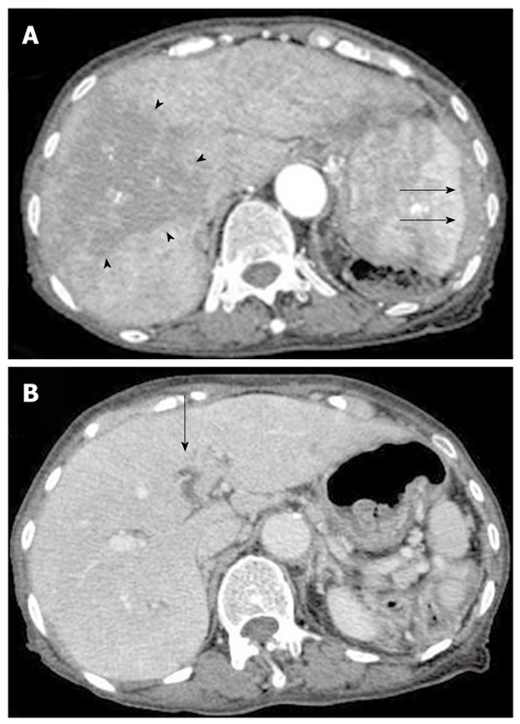
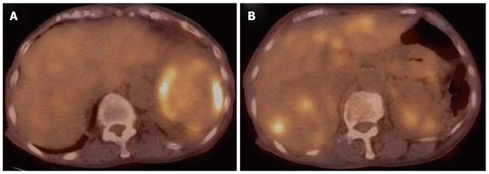
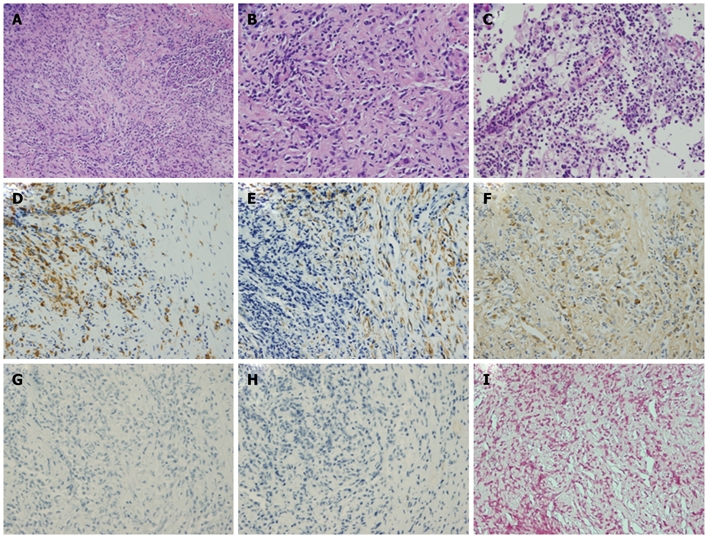
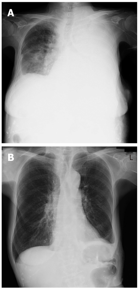
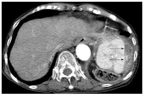
An inflammatory pseudotumor (IPT) is a relatively rare lesion characterized by chronic infiltration of inflammatory cells and areas of fibrosis. IPTs are difficult to diagnose because of the absence of specific symptoms or of characteristic hematological or radiological findings. In this study, a case of a woman aged over 70 years was reported, who presented with a general malaise lasting more than two months. A computed tomography scan demonstrated a diffusely spread lesion of the liver with a portal vein occlusion and a splenic lesion surrounded by a soft density layer. Since the percutaneous liver biopsy showed findings that suggested an IPT, although the radiological findings did not exclude the possibility of a malignancy, we performed a percutaneous spleen biopsy to enable a more definitive diagnosis. The microscopic findings from the spleen specimen lead us to a diagnosis of IPT involving the liver and spleen. Subsequent steroid pulse therapy was effective, and rapid resolution of the disease was observed.
Keywords: Inflammatory pseudotumor; Percutaneous liver biopsy; Percutaneous spleen biopsy; Steroid pulse therapy.
Figures
References
-
- Brunn H. Two interesting benign lung tumors of contradictory histopathology. Remarks on the necessity for maintaining the chest tumor registry. J Thorac Cardiovasc Surg. 1939;9:119–131.
-
- Cotelingam JD, Jaffe ES. Inflammatory pseudotumor of the spleen. Am J Surg Pathol. 1984;8:375–380. - PubMed
-
- Nam KJ, Kang HK, Lim JH. Inflammatory pseudotumor of the liver: CT and sonographic findings. AJR Am J Roentgenol. 1996;167:485–487. - PubMed
-
- Lupovitch A, Chen R, Mishra S. Inflammatory pseudotumor of the liver. Report of the fine needle aspiration cytologic findings in a case initially misdiagnosed as malignant. Acta Cytol. 1989;33:259–262. - PubMed
Publication types
MeSH terms
LinkOut - more resources
Full Text Sources
Medical
