

Near-IR Multi-modal Imaging of Natural Occlusal Lesions
- PMID: 22228979
- PMCID: PMC3251260
- DOI: 10.1117/12.816866
Near-IR Multi-modal Imaging of Natural Occlusal Lesions
Abstract
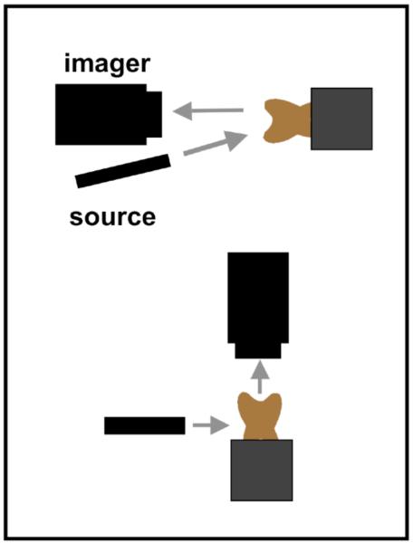
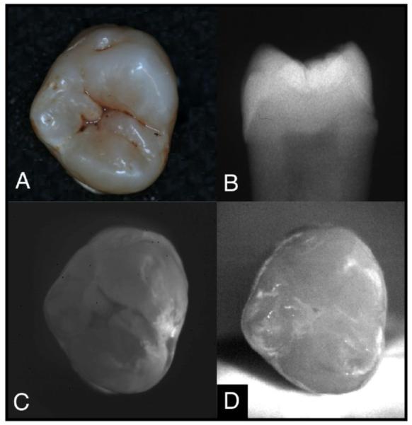
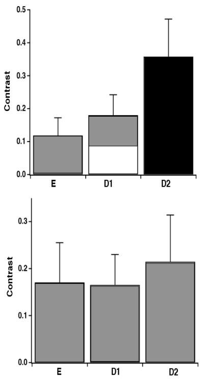
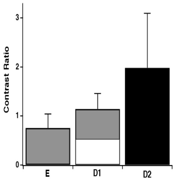
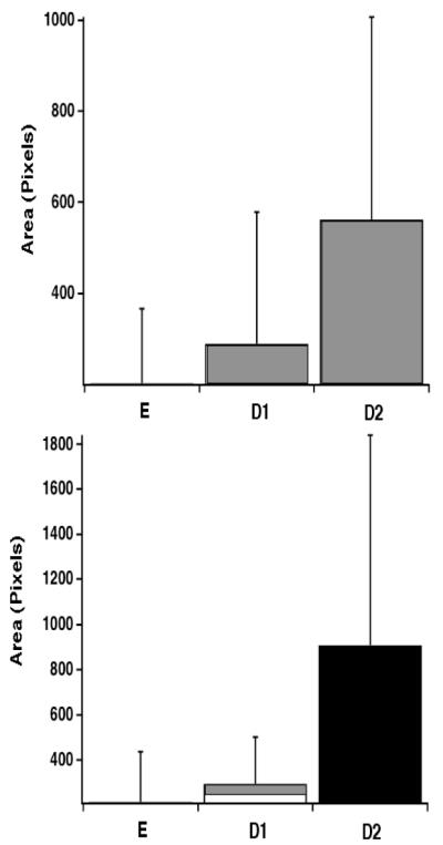
Reflectance and transillumination imaging show demineralization with high contrast in the near-IR. The objective of this study is to use lesion size and contrast acquired in reflectance and transillumination near-infrared imaging modes to estimate the severity of natural occlusal caries lesions. Previous studies have shown that near-infrared (NIR) light can be used to effectively image artificial carious lesions. However, its efficacy on natural lesions requires further exploration. Fifty extracted teeth with varying amounts of occlusal decay were examined using a NIR imaging system operating at 1310-nm. Image analysis software was used to calculate contrast values between sound and carious tooth structure. After imaging, teeth were histologically sampled at 1-mm intervals in order to determine lesion depth. Lesion contrast in transillumination mode significantly increased with lesion depth (p<0.001), while lesion contrast in reflectance mode did not increase. The lesion area demonstrated a significant increase with lesion severity in both imaging modes. These results suggest that lesion contrast and area can be used to estimate lesion severity in NIR images.
Figures
Similar articles
-
Transillumination and optical coherence tomography for the detection and diagnosis of enamel caries.Cochrane Database Syst Rev. 2021 Jan 27;1(1):CD013855. doi: 10.1002/14651858.CD013855. Cochrane Database Syst Rev. 2021. PMID: 33502759 Free PMC article.
-
A system for simultaneous near-infrared reflectance and transillumination imaging of occlusal carious lesions.Proc SPIE Int Soc Opt Eng. 2016 Feb 13;9692:96920A. doi: 10.1117/12.2218656. Epub 2016 Feb 29. Proc SPIE Int Soc Opt Eng. 2016. PMID: 27006524 Free PMC article.
-
Multispectral near-infrared reflectance and transillumination imaging of occlusal carious lesions: Variation in lesion contrast with lesion depth.Proc SPIE Int Soc Opt Eng. 2018 Jan-Feb;10473:1047305. doi: 10.1117/12.2296013. Epub 2018 Feb 8. Proc SPIE Int Soc Opt Eng. 2018. PMID: 29497229 Free PMC article.
-
Multispectral near-IR reflectance and transillumination imaging of teeth.Biomed Opt Express. 2011 Oct 1;2(10):2804-14. doi: 10.1364/BOE.2.002804. Epub 2011 Sep 15. Biomed Opt Express. 2011. PMID: 22025986 Free PMC article.
-
Multispectral near-IR reflectance imaging of simulated early occlusal lesions: variation of lesion contrast with lesion depth and severity.Lasers Surg Med. 2014 Mar;46(3):203-15. doi: 10.1002/lsm.22216. Epub 2013 Dec 27. Lasers Surg Med. 2014. PMID: 24375543 Free PMC article.
Cited by
-
Assessment of remineralization in simulated enamel lesions via dehydration with near-IR reflectance imaging.Proc SPIE Int Soc Opt Eng. 2015 Feb 24;9306:93060H. doi: 10.1117/12.2083655. Proc SPIE Int Soc Opt Eng. 2015. PMID: 25914494 Free PMC article.
-
Influence of stains on lesion contrast in the pits and fissures of tooth occlusal surfaces from 800-1600-nm.Proc SPIE Int Soc Opt Eng. 2016 Feb 13;9692:96920X. doi: 10.1117/12.2218663. Epub 2016 Feb 29. Proc SPIE Int Soc Opt Eng. 2016. PMID: 26997740 Free PMC article.
-
Imaging Natural Occlusal Caries Lesions with Optical Coherence Tomography.Proc SPIE Int Soc Opt Eng. 2010;7549:75490N. doi: 10.1117/12.849344. Proc SPIE Int Soc Opt Eng. 2010. PMID: 22228981 Free PMC article.
-
Transillumination and optical coherence tomography for the detection and diagnosis of enamel caries.Cochrane Database Syst Rev. 2021 Jan 27;1(1):CD013855. doi: 10.1002/14651858.CD013855. Cochrane Database Syst Rev. 2021. PMID: 33502759 Free PMC article.
-
Detection, Diagnosis, and Monitoring of Early Caries: The Future of Individualized Dental Care.Diagnostics (Basel). 2023 Dec 12;13(24):3649. doi: 10.3390/diagnostics13243649. Diagnostics (Basel). 2023. PMID: 38132233 Free PMC article. Review.
References
-
- Barenie J, Leske G, Ripa LW. The use of fiber optic transillumination for the detection of proximal caries. Oral Surg. 1973;36:891–897. - PubMed
-
- Pine CM. Fiber-Optic Transillumination (FOTI) in Caries Diagnosis, Early Detection of dental caries. Indiana University; 1996. pp. 51–66.
-
- Peltola J, Wolf J. Fiber optics transillumination in caries diagnosis. Proc Finn Dent Soc. 1981;77:240–244. - PubMed
-
- Holt RD, Azeevedo MR. Fiber Optic transillumination and radiographs in diagnosis of approximal caries in primary teeth. Community Dent Health. 1989;6:239–247. - PubMed
-
- Mitropoulis CM. The use of fiber optic transillumination in the diagnosis of posterior approximal caries in clinical trials. Caries Res. 1985;19:379–384. - PubMed
Grants and funding
LinkOut - more resources
Full Text Sources
Other Literature Sources
Miscellaneous