

Vascular network modeling reveals significant differences in vascular morphology in growth-restricted placentas
- PMID: 22229062
- PMCID: PMC3252882
Vascular network modeling reveals significant differences in vascular morphology in growth-restricted placentas
Abstract
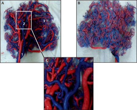
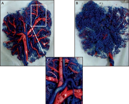
Aim: To construct and examine models of the vascular networks using the technique of vascular corrosion casting in placentas collected from normal pregnancies and from pregnancies complicated by fetal growth restriction (FGR).
Methods: Twenty placentas were collected from normal term pregnancies (Group NP) and an equal number from pregnancies with idiopathic term FGR (Group FGR) and placental vascular network models constructed by perfusing an acrylic-based solution separately into the umbilical vein and arteries. Placental blood volumes and blood vessel characteristics (number of branches, diameter, and morphology) were then examined and compared.
Results: In placentas from Group NP, the veins branched five to seven times with a peripheral artery-to-vein ratio ranging from 1:2 to 1:3. In placentas from Group FGR, the veins branched only four to five times with an artery-to-vein ratio of 1:1 to 2:1 and increased evidence of nodularity and pitting of the vessel walls. The two groups showed significant differences in placental blood volume and in the mean diameters of umbilical veins and arteries. In Group FGR, significant positive correlations could be found between birth weight and placental volume, venous diameters, and select arterial diameters.
Conclusion: Vascular network models can be constructed from term placentas. Such modeling may provide novel insights and improve our understanding of the placental vascular system in both health and disease.
Keywords: Fetal growth restriction; Placenta; Pregnancy; Vascular corrosion casting.
Figures
References
-
- Biswas S, Ghosh SK. Gross morphological changes of placentas associated with intrauterine growth restriction of fetuses: a case control study. Early Hum Dev. 2008;84:357–362. - PubMed
-
- Sağol S, Ozkinay E, Oztekin K, Ozdemir N. The comparison of uterine artery Doppler velocimetry with the histopathology of the placental bed. Aust N Z J Obstet Gynaecol. 1999;39:324–329. - PubMed
-
- Verli FD, Rossi-Schneider TR, Schneider FL, et al. Vascular corrosion casting technique steps. Scanning. 2007;29:128–132. - PubMed
-
- Tang L, Chung MS, Liu Q, Shin DS. Advanced features of whole body sectioned images: Virtual Chinese Human. Clin Anat. 2010;23:523–529. - PubMed
-
- Chen CL, Guo HX, Liu P, et al. Three-dimensional reconstruction of the uterine vascular supply through vascular casting and thin slice computed tomography scanning. Minim Invasive Ther Allied Technol. 2009;18:98–102. - PubMed
LinkOut - more resources
Full Text Sources
Miscellaneous