

Dynamic contrast-enhanced MR imaging findings of bone metastasis in patients with prostate cancer
- PMID: 22229077
- PMCID: PMC3252556
- DOI: 10.4329/wjr.v3.i10.241
Dynamic contrast-enhanced MR imaging findings of bone metastasis in patients with prostate cancer
Abstract
Aim: To evaluate the dynamic contrast-enhanced magnetic resonance imaging (DCE-MRI) findings of bone metastasis in prostate cancer patients.
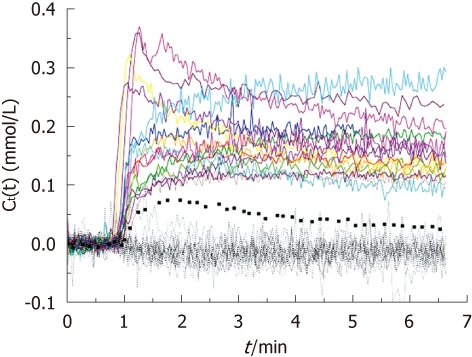
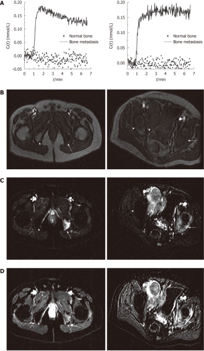
Methods: Sixteen men with a diagnosis of metastatic prostate cancer to bones were examined with DCE-MRI at 1.5 Tesla. The mean contrast agent concentration vs time curves for bone metastasis and normal bone were calculated and K(trans) and ve values were estimated and compared.
Results: An early significant enhancement (wash-out: n = 6, plateau: n = 8 and persistent: n = 2) was detected in all bone metastases (n = 16). Bone metastasis from prostate cancer showed significant enhancement and high K(trans) and ve values compared to normal bone which does not enhance in the elderly population. The mean K(trans) was 0.101/min and 0.0051/min (P < 0.001), the mean ve was 0.141 and 0.0038 (P < 0.001), for bone metastases and normal bone, respectively.
Conclusion: DCE-MRI and its quantitative perfusion parameters may have a role in improving the detection of skeletal metastasis in prostate cancer patients.
Keywords: Bone; Cancer; Dynamic contrast-enhanced MR imaging; Metastasis; Prostate.
Figures
References
-
- Jemal A, Siegel R, Ward E, Hao Y, Xu J, Thun MJ. Cancer statistics, 2009. CA Cancer J Clin. 2009;59:225–249. - PubMed
-
- Manyak MJ, Javitt MC. The role of computerized tomography, magnetic resonance imaging, bone scan, and monoclonal antibody nuclear scan for prognosis prediction in prostate cancer. Semin Urol Oncol. 1998;16:145–152. - PubMed
-
- Freedman GM, Negendank WG, Hudes GR, Shaer AH, Hanks GE. Preliminary results of a bone marrow magnetic resonance imaging protocol for patients with high-risk prostate cancer. Urology. 1999;54:118–123. - PubMed
-
- Venkitaraman R, Sohaib SA, Barbachano Y, Parker CC, Khoo V, Huddart RA, Horwich A, Dearnaley DP. Detection of occult spinal cord compression with magnetic resonance imaging of the spine. Clin Oncol (R Coll Radiol) 2007;19:528–531. - PubMed
-
- Lecouvet FE, Geukens D, Stainier A, Jamar F, Jamart J, d’Othée BJ, Therasse P, Vande Berg B, Tombal B. Magnetic resonance imaging of the axial skeleton for detecting bone metastases in patients with high-risk prostate cancer: diagnostic and cost-effectiveness and comparison with current detection strategies. J Clin Oncol. 2007;25:3281–3287. - PubMed
Grants and funding
LinkOut - more resources
Full Text Sources
