

Theranostic applications: Non-ionizing cellular and molecular imaging through innovative nanosystems for early diagnosis and therapy
- PMID: 22229079
- PMCID: PMC3252558
- DOI: 10.4329/wjr.v3.i10.49
Theranostic applications: Non-ionizing cellular and molecular imaging through innovative nanosystems for early diagnosis and therapy
Abstract
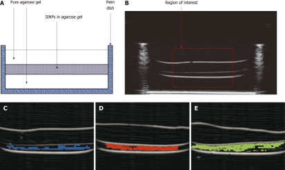
Modern medicine is expanding the possibilities of receiving "personalized" diagnosis and therapies, providing minimal invasiveness, technological solutions based on non-ionizing radiation, early detection of pathologies with the main objectives of being operator independent and with low cost to society. Our research activities aim to strongly contribute to these trends by improving the capabilities of current diagnostic imaging systems, which are of key importance in possibly providing both optimal diagnosis and therapies to patients. In medical diagnostics, cellular imaging aims to develop new methods and technologies for the detection of specific metabolic processes in living organisms, in order to accurately identify and discriminate normal from pathological tissues. In fact, most diseases have a "molecular basis" that detected through these new diagnostic methodologies can provide enormous benefits to medicine. Nowadays, this possibility is mainly related to the use of Positron Emission Tomography, with an exposure to ionizing radiation for patients and operators and with extremely high medical diagnostics costs. The future possible development of non-ionizing cellular imaging based on techniques such as Nuclear Magnetic Resonance or Ultrasound, would represent an important step towards modern and personalized therapies. During the last decade, the field of nanotechnology has made important progress and a wide range of organic and inorganic nanomaterials are now available with an incredible number of further combinations with other compounds for cellular targeting. The availability of these new advanced nanosystems allows new scenarios in diagnostic methodologies which are potentially capable of providing morphological and functional information together with metabolic and cellular indications.
Keywords: Intelligent nanosystems for cellular targeting; Magnetic resonance and ultrasound; Molecular imaging; Non-ionizing diagnostic techniques; Personalized medicine in the oncological and vascular field.
Figures

References
-
- Ferlay J, Shin HR, Bray F, Forman D, Mathers C, Parkin DM. Estimates of worldwide burden of cancer in 2008: GLOBOCAN 2008. Int J Cancer. 2010;127:2893–2917. - PubMed
-
- Albreht T, McKee M, Alexe DM, Coleman MP, Martin-Moreno JM. Making progress against cancer in Europe in 2008. Eur J Cancer. 2008;44:1451–1456. - PubMed
-
- Casciaro S, Thome JR. Thermal performance of flooded evaporators, part 1: review of boiling heat transfer studies. ASHRAE Trans. 2001;107:903–918.
LinkOut - more resources
Full Text Sources
