. 2012 Jun 15;16(12):1421-33.
doi: 10.1089/ars.2011.4173. Epub 2012 Feb 28.

Mitochondrion-derived reactive oxygen species lead to enhanced amyloid beta formation

Affiliations

Mitochondrion-derived reactive oxygen species lead to enhanced amyloid beta formation

Kristina Leuner et al. Antioxid Redox Signal. .

Abstract

Aims: Intracellular amyloid beta (Aβ) oligomers and extracellular Aβ plaques are key players in the progression of sporadic Alzheimer's disease (AD). Still, the molecular signals triggering Aβ production are largely unclear. We asked whether mitochondrion-derived reactive oxygen species (ROS) are sufficient to increase Aβ generation and thereby initiate a vicious cycle further impairing mitochondrial function.

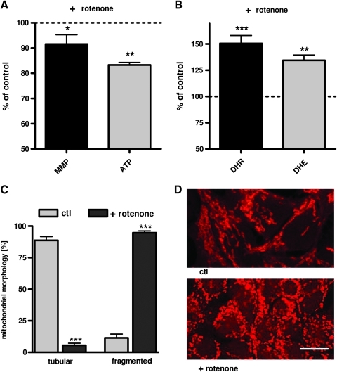
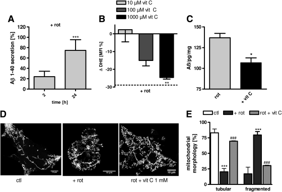
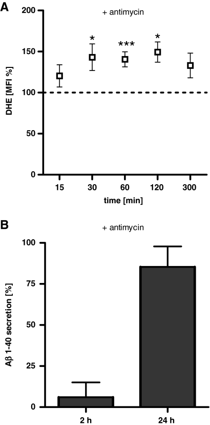
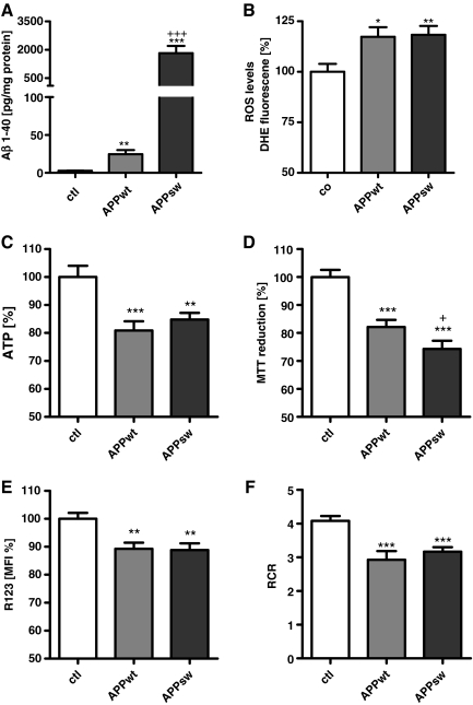
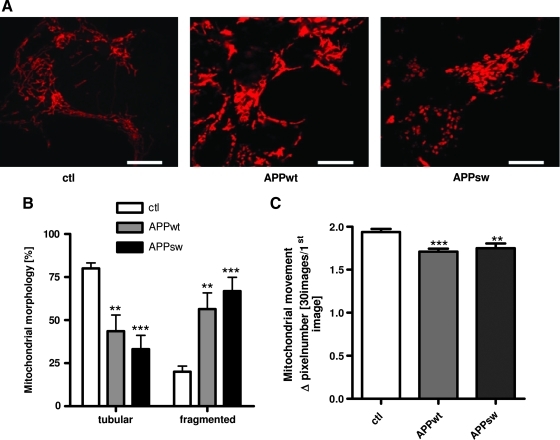
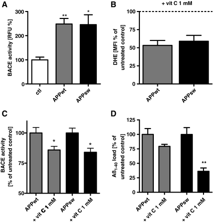
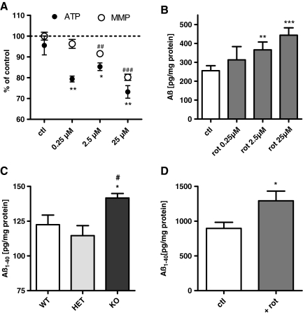
Results: Complex I and III dysfunction was induced in a cell model using the respiratory inhibitors rotenone and antimycin, resulting in mitochondrial dysfunction and enhanced ROS levels. Both treatments lead to elevated levels of Aβ. Presence of an antioxidant rescued mitochondrial function and reduced formation of Aβ, demonstrating that the observed effects depended on ROS. Conversely, cells overproducing Aβ showed impairment of mitochondrial function such as comprised mitochondrial respiration, strongly altered morphology, and reduced intracellular mobility of mitochondria. Again, the capability of these cells to generate Aβ was partly reduced by an antioxidant, indicating that Aβ formation was also ROS dependent. Moreover, mice with a genetic defect in complex I, or AD mice treated with a complex I inhibitor, showed enhanced Aβ levels in vivo.

Innovation: We show for the first time that mitochondrion-derived ROS are sufficient to trigger Aβ production in vitro and in vivo.

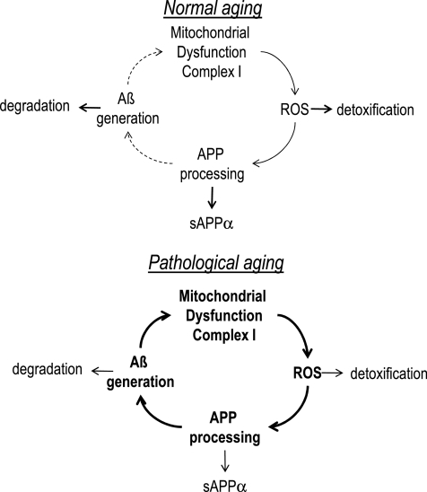
Conclusion: Several lines of evidence show that mitochondrion-derived ROS result in enhanced amyloidogenic amyloid precursor protein processing, and that Aβ itself leads to mitochondrial dysfunction and increased ROS levels. We propose that starting from mitochondrial dysfunction a vicious cycle is triggered that contributes to the pathogenesis of sporadic AD.


Figures

FIG. 1.
FIG. 1.
Complex I inhibitor rotenone leads to mitochondrial dysfunction, fragmentation, and enhanced reactive oxygen species (ROS) production. (A) Mitochondrial membrane potential (MMP; R123 mean fluorescence intensity [MFI] normalized on% of untreated control) and ATP levels were significantly reduced after 24-h treatment with rotenone (25 μM). (B) Levels of superoxide anion radicals (dihydroethidium [DHE] fluorescence normalized on% of untreated controls), and cytosolic ROS (dihydrorhodamin [DHR] fluorescence protein normalized on% of untreated controls) were increased after 2 h treatment with rotenone (25 μM). (C) Histogram of quantitative analysis of mitochondrial morphology (Mitotracker CMXRos) after 24 h rotenone insult (100 mitochondria/n). (D) Representative confocal images that revealed changes in the mitochondrial morphology of human embryonic kidney (HEK) cells treated with rotenone (25 μM, 24 h; bars represent 10 μm) compared to untreated controls after mitochondrial staining with MitoTracker CMXRos (red). (A–C) n=6±standard error of the mean (SEM); unpaired t-test; *p<0.05, **p<0.01, ***p<0.001. (To see this illustration in color the reader is referred to the web version of this article at www.liebertonline.com/ars).
FIG. 2.
FIG. 2.
Complex I dysfunction leads to ROS-dependent Aβ generation. (A) Soluble Aβ1-40 levels in HEK cells are significantly increased after 24-h insult with rotenone (rot; 25 μM) compared to control cells (ctl). (B) 4-h preincubation with vitamin C (1000 μM) leads to a significant reduction of superoxide anion radicals (Δ DHE MFI, normalized on % of untreated controls) after rotenone insult (25 μM, 2 h). (C) ROS scavenging using vitamin C (1000 μM) results in significantly reduced Aβ1-40 levels after 24 h insult with rotenone (25 μM). (D) Representative images reveal changes in mitochondrial morphology when cells are pretreated with vitamin C (1000 μM) and afterward stressed with rotenone (25 μM) compared to untreated controls. Mitochondria were stained with MitoTracker CMXRos (bars represent 10 μm). (E) Histogram of quantitative analysis of mitochondrial morphology in the presence and absence of vitamin C (1000 μM, preincubation 4 h) after 24-h rotenone insult (100 mitochondria/n). (A–C) n=6±SEM; unpaired t-test; *p<0.05, **p<0.01, ***p<0.001. (E) n=6±SEM; unpaired t-test, ***p<0.001 ctl compared to rotenone treated cells, ###p<0.001 rotenone treated cells against vit C+rotenone-treated cells.
FIG. 3.
FIG. 3.
Complex III dysfunction also leads to enhanced ROS and Aβ levels. (A) Levels of superoxide anion radicals (DHE MFI normalized on untreated controls) were significantly increased after the treatment with antimycin (100 μM) for 30, 60, and 120 min. (B) Soluble Aβ1-40 levels in HEK cells are significantly increased after 24-h insult with antimycin (100 μM) compared to cells without antimycin. (A, B) n=6±SEM; unpaired t-test; *p<0.05, ***p<0.001.
FIG. 4.
FIG. 4.
Mitochondrial function is greatly impaired in wild-type amyloid precursor protein (APPwt) and APP containing the Swedish mutation (APPsw) cells. (A)1-40 levels in transfected HEK cells are significantly increased in APPwt and APPsw cells. (B) Superoxide anion radicals (DHE fluorescence units/mg protein normalized on control cells) are significantly enhanced in APPwt and APPsw cells. (C) ATP levels and cell viability (D) are reduced in both APPwt and APPsw. (E) MMP (R123 units/mg protein expressed as MFI normalized to control cells) is significantly impaired in APPwt and APPsw cells. (F) Using a high-resolution respiratory system, respiratory control ratio (RCR; basal respiration/respiration under the treatment with oligomycin) representing the mitochondrial coupling state is significantly reduced again in APPwt and APPsw cells (A–E) n=6±SEM; unpaired t-test; ctl against APPwt or APPsw *p<0.05, **p<0.01, ***p<0.001. +p<0.05 APPwt against APPsw, +++p<0.001 APPwt against APPsw.
FIG. 5.
FIG. 5.
Mitochondrial morphology is strongly altered in APPwt and APPsw cells. (A) Representative example of confocal microscopic images revealing mitochondrial morphology changes in APPwt and APPsw cells. Mitochondrial staining was done using MitoTracker CMXRos (red, bars represent 10 μm). (B) Histogram of mitochondrial morphology quantification (100 mitochondria/n were analyzed). (C) Live cell imaging of mitochondria stained with MitoTracker Deep Red. Images were recorded over 2 min (one picture every 1.5 s) to study mitochondrial dynamics. Histogram depicting quantitative changes in mitochondrial dynamics in APPwt and APPsw cells. (B, C) n=6±SEM; unpaired t-test; **p<0.01, ***p<0.001 ctl against APPwt or ctl against APPsw, respectively. (To see this illustration in color the reader is referred to the web version of this article at www.liebertonline.com/ars).
FIG. 6.
FIG. 6.
ROS are also involved in amyloidogenic APP processing in APPwt and APPsw cells. (A) Basal β-site APP cleaving enzyme 1 (BACE1) activity is increased in APPwt and APPsw cells. (B) Effectivity of ROS scavenging in APPwt and APPsw cells by vitamin C on DHE MFI (1000 μM; 4 h incubation). (C) BACE1 activity is reduced in APPwt and APPsw cells by 4-h incubation of vitamin C (1000 μM). (D) Reduction of Aβ levels in APPwt and APPsw cells by vitamin C (1000 μM, 4 h incubation). (A–D) n=6±SEM; unpaired t-test; *p<0.05, **p<0.01. (A) ctl against APPwt or ctl against APPsw. (C, D) APPwt or APPsw against APPwt+vit C or APPsw against APPsw+vit C.
FIG. 7.
FIG. 7.
Enhanced Aβ levels in a neuronal cell model and two animal models of complex I dysfunction. (A) SH-SY5Y cells were treated with rotenone (0.25, 2.5, and 25 μM) for 24 h and ATP and MMP as well as soluble Aβ1-40 levels (B) were determined n=6±SEM, unpaired t-test, (A) *p<0.05, **p<0.01, ATP ctl against rot, ##p<0.01, ###p<0.001 MMP against rot. (B) **p<0.01 ctl against rot 2.5 μM, ***p<0.001 ctl against rot 25 μM. (C) APP transgenic animals received rotenone via i.p. application (10 mg/kg/body weight) or vehicle for 3 days and soluble Aβ1-40 levels in brain homogenates were measured. n=6±SEM per treatment group unpaired t-test *p<0.05. (D) In homozygous knockout (KO) Ndufs4 mice, Aβ1-40 levels are significantly increased compared to wild-type (wt) and heterozygous (HET) mice. Ndufs4 mice wt n=5±SEM, HET n=5±SEM, KO n=12±SEM; unpaired t-test; *p<0.05 wt against KO, #p<0.05 HET against KO.
FIG. 8.
FIG. 8.
Schematic illustration of mitochondrial dysfunction in healthy brain aging and pathological brain aging. In normal brain aging there is an equilibrium between mitochondrion-derived ROS, APP processing, and Aβ generation on the one hand and detoxification and degradation mechanisms on the other hand. During the disease process this balance is shifted toward the toxic mechanism with pre-ponderance for mitochondrion-derived ROS, APP processing associated with increase BACE activity, and Aβ generation.

References

    1. Anandatheerthavarada HK. Devi L. Amyloid precursor protein and mitochondrial dysfunction in Alzheimer's disease. Neuroscientist. 2007;13:626–638. - PubMed
    1. Andreyev AI. Kushnareva YE. Starkov AA. Mitochondrial metabolism of reactive oxygen species. Biochem Mosc. 2005;70:200–214. - PubMed
    1. Behl C. Apoptosis and Alzheimer's disease. J Neural Transm. 2000;107:1325–1344. - PubMed
    1. Benard G. Bellance N. James D. Parrone P. Fernandez H. Letellier T. Rossignol R. Mitochondrial bioenergetics and structural network organization. J Cell Sci. 2007;120:838–848. - PubMed
    1. Bishop NA. Lu T. Yankner BA. Neural mechanisms of ageing and cognitive decline. Nature. 2010;464:529–535. - PMC - PubMed

Publication types

MeSH terms

LinkOut - more resources