. 2012 Feb;22(1):58-68.
doi: 10.1089/nat.2011.0316. Epub 2012 Jan 9.

Degradation of nuclease-stabilized RNA oligonucleotides in Mycoplasma-contaminated cell culture media

Affiliations

Degradation of nuclease-stabilized RNA oligonucleotides in Mycoplasma-contaminated cell culture media

Frank J Hernandez et al. Nucleic Acid Ther. 2012 Feb.

Abstract

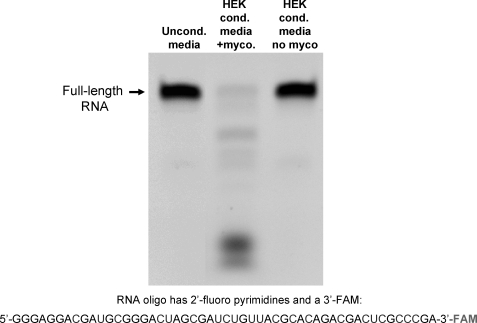
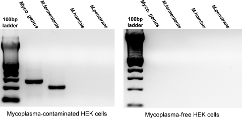
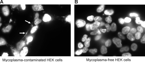
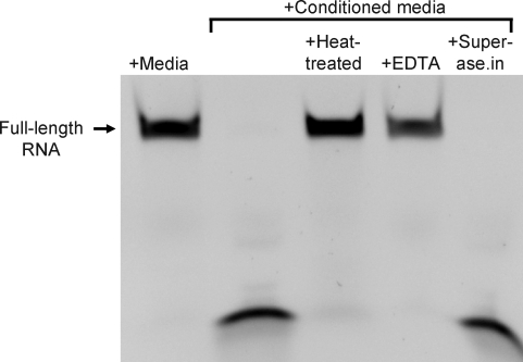
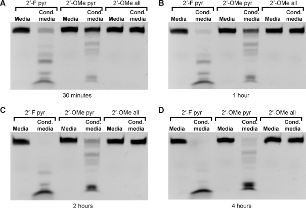
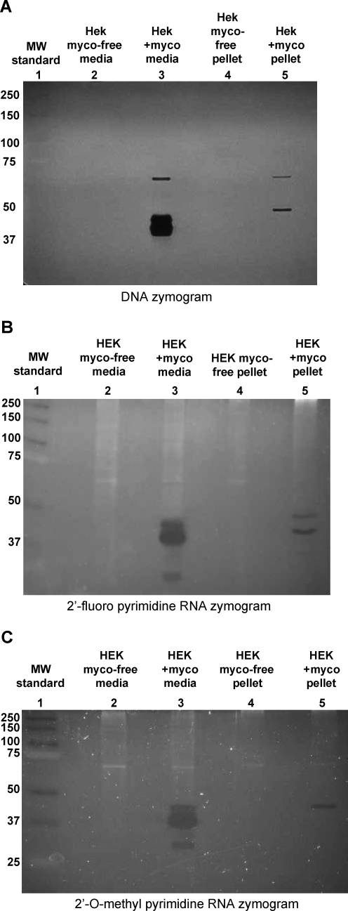
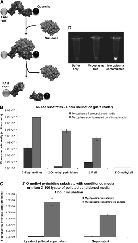
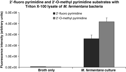
Artificial RNA reagents such as small interfering RNAs (siRNAs) and aptamers often must be chemically modified for optimal effectiveness in environments that include ribonucleases. Mycoplasmas are common bacterial contaminants of mammalian cell cultures that are known to produce ribonucleases. Here we describe the rapid degradation of nuclease-stabilized RNA oligonucleotides in a human embryonic kidney 293 (HEK) cell culture contaminated with Mycoplasma fermentans, a common species of mycoplasma. RNA with 2'-fluoro- or 2'-O-methyl- modified pyrimidines was readily degraded in conditioned media from this culture, but was stable in conditioned media from uncontaminated HEK cells. RNA completely modified with 2'-O-methyls was not degraded in the mycoplasma-contaminated media. RNA zymogram analysis of conditioned culture media and material centrifuged from the media revealed several distinct protein bands (ranging from 30 to 68 kDa) capable of degrading RNA with 2'-fluoro- or 2'-O-methyl-modified pyrimidines. Finally, the mycoplasma-associated nuclease was detected in material centrifuged from the contaminated culture supernatants in as little as 15 minutes with an RNA oligo-containing 2'-O-methyl-modified pyrimidines and labeled with a 5'-fluorescein amidite (FAM) and 3'-quencher. These results suggest that mycoplasma contamination may be a critical confounding variable for cell culture experiments involving RNA-based reagents, with particular relevance for applications involving naked RNA (e.g., aptamer-siRNA chimeras).


Figures

FIG. 1.
FIG. 1.
An RNA oligo with 2′-fluoro-modified pyrimidines is degraded in conditioned media from mycoplasma contaminated HEK cells. A 51 nucleotide-long RNA oligo, composed of 2′-fluoro modified pyrimidines and unmodified purines was incubated with unconditioned culture media, culture media conditioned by mycoplasma contaminated HEK cells, or culture media from uncontaminated HEK cells for 4 hours at 37°C. The RNA was then resolved on a urea-acrylamide denaturing gel and imaged with a digital camera and UV light transillumination; the oligo is labeled on its 3′ end with FAM.
FIG. 2.
FIG. 2.
Polymerase chain reaction (PCR)-based assay detected mycoplasma contamination in HEK cell culture and identified species as M. fermentans. PCR primer sets specific for genomic components of M. fermentans, Mycoplasma hominis, Mycoplasma penetrans, or for a region of the mycoplasma genome that is conserved within the genus were used to amplify DNA present in conditioned culture media. Expected PCR product sizes are as follows: mycoplasma genus PCR: 280 bp; M. fermentans: 206 bp; M. hominis: 170 bp; M. penetrans: 470 bp. Material centrifuged from one HEK cell culture was used as template for PCRs run on the gel on the left, while material centrifuged from a second HEK cell culture recently obtained from ATCC was used as template for PCRs run on the gel on the right.
FIG. 3.
FIG. 3.
Mycoplasma contamination detected in HEK cells with DAPI (4′,6-diamidino-2-phenylindole, dihydrochloride) stain. HEK cells from 2 cultures—one older culture (A) and one recently obtained from ATCC (B)—were plated on glass-bottomed culture dishes and stained with the nucleic acid stain DAPI. Note the extranuclear punctate pattern of fluorescence (arrows in panel A), a telltale sign of mycoplasma contamination, found in the older culture.
FIG. 4.
FIG. 4.
Nuclease activity in mycoplasma-contaminated culture media is sensitive to heat treatment and depends on the presence of divalent cations. A 51-nucleotide-long RNA oligo, composed of 2′-fluoro modified pyrimidines and unmodified purines was incubated for 4 hours at 37°C, either in fresh culture media or culture media conditioned by mycoplasma contaminated HEK cells. To determine sensitivity of the nuclease activity to heat, conditioned media was heated to 95°C for 10 minutes prior to combination and incubation with RNA. To determine the dependence of the nuclease activity on divalent cations, conditioned media was incubated with RNA in the presence of 10 mM EDTA. A broad-spectrum RNase inhbitor, Superase.in, was coincubated (at 1 U/μL) with the RNA in conditioned media to determine the sensitivity of the nuclease activity to this reagent. The RNA was resolved on a urea-acrylamide denaturing gel and imaged with a digital camera and UV light transillumination; the oligo is labeled on its 3′ end with FAM.
FIG. 5.
FIG. 5.
Time-course and substrate specificity of nuclease activity in mycoplasma contaminated culture media. Three 51-nucleotide RNA oligos of identical sequence but with different chemical modifications (2′-fluoro modified pyrimidines only, 2′-O-methyl modified pyrimidines only, or complete 2′-O-methyl modification) were incubated with fresh culture media or with culture media conditioned by mycoplasma contaminated HEK cells at 37°C for 30 minutes (A), 1 hour (B), 2 hours (C), and 4 hours (D). The RNA was resolved on urea-acrylamide denaturing gels and imaged with a digital camera and UV light transillumination; the oligos are labeled on their 3′ ends with FAM.
FIG. 6.
FIG. 6.
Nuclease sizes determined with zymograms. Concentrated media or material pelleted by centrifugation from media conditioned by mycoplasma-free or mycoplasma-contaminated HEK cells was resolved on 8% acrylamide-sodium dodecyl sulfate (SDS) gels embedded with DNA (A), RNA with 2′-fluoro-modified pyrimidines (B), or RNA with 2′-O-methyl-modified pyrimidines (C). After running, the gels were washed with SDS-free buffer containing divalent cations and incubated at 37°C for 2 hours to allow nuclease digestion. The gels were stained with ethidium bromide (DNA zymogram) or SYBR Gold nucleic acid gel stain and imaged with a digital camera and UV light transillumination, revealing protein bands with nuclease activity.
FIG. 7.
FIG. 7.
Rapid detection of mycoplasma-associated nuclease activity with chemically modified RNase substrates. (A) The basis for nuclease detection with RNase substrates is illustrated. RNA oligonucleotides (5′-UCUCGUACGUUC-3′) with chemically modified nucleotides, labeled on the 5′ ends with fluorescein amidite (FAM) are not fluorescent due to the close proximity of a 3′-quencher to the FAM. Upon degradation of the oligo, the quencher diffuses away from the FAM and the FAM exhibits green fluorescence. (B) Mycoplasma-associated nuclease activity is detected with various RNase substrates. RNase substrates with the chemically modified RNA compositions indicated were coincubated with culture media conditioned by mycoplasma-free or mycoplasma-contaminated human embryonic kidney (HEK) cells for 4 hours at 37°C. Fluorescence of these reactions was then measured with a fluorescence plate reader. Background fluorescence levels determined by the fluorescence level of each RNase substrate incubated in serum-free unconditioned media have been subtracted from each experimental value. (C) The RNase substrate with 2-O-methyl-modified pyrimidines was incubated with the culture supernatant or a lysate prepared from material centrifuged from the supernatants of mycoplasma-free or mycoplasma-contaminated HEK cells. This assay was carried out as described for (B), above, except that the incubation was for only 1 hour. (D) The RNase substrate with 2′-O-methyl-modified pyrimidines was incubated for 15 minutes with a lysate prepared from material centrifuged from the supernatants of mycoplasma-free or mycoplasma-contaminated HEK cells. Eppendorf tubes with these reactions were imaged with a digital camera and ultraviolet (UV) light transillumination.
FIG. 8.
FIG. 8.
Nuclease activity in a Mycoplasma fermentans lysate. RNase substrates with the indicated compositions were incubated with a lysate of M. fermentans bacteria for 1 hour at 37°C, and fluorescence levels were measured with a fluorescence plate reader. A mock lysate prepared from centrifuged, bacteria-free broth served as negative control. Background fluorescence levels (determined by measuring the fluorescence level of each RNase substrate incubated in phosphate buffered saline have been subtracted from each experimental value.

Similar articles

Cited by

References

    1. ALLERSON C.R. SIOUFI N. JARRES R. PRAKASH T.P. NAIK N. BERDEJA A. WANDERS L. GRIFFEY R.H. SWAYZE E.E. BHAT B. Fully 2′-modified oligonucleotide duplexes with improved in vitro potency and stability compared to unmodified small interfering RNA. J. Med. Chem. 2005;48:901–904. - PubMed
    1. BEHLKE M.A. Chemical modification of siRNAs for in vivo use. Oligonucleotides. 2008;18:305–319. - PubMed
    1. BENDJENNAT M. BLANCHARD A. LOUTFI M. MONTAGNIER L. BAHRAOUI E. Purification and characterization of Mycoplasma penetrans Ca2+/Mg2+-dependent endonuclease. J. Bacteriol. 1997;179:2210–2220. - PMC - PubMed
    1. BOERLIN P. KUHNERT P. HUSSY D. SCHAELLIBAUM M. Methods for identification of Staphylococcus aureus isolates in cases of bovine mastitis. J. Clin. Microbiol. 2003;41:767–771. - PMC - PubMed
    1. CERCHIA L. DUCONGE F. PESTOURIE C. BOULAY J. AISSOUNI Y. GOMBERT K. TAVITIAN B. DE FRANCISCIS V. LIBRI D. Neutralizing aptamers from whole-cell SELEX inhibit the RET receptor tyrosine kinase. PLoS Biol. 2005;3:e123. - PMC - PubMed

Publication types

LinkOut - more resources