. 2012 Dec;17(12):1261-71.
doi: 10.1038/mp.2011.180. Epub 2012 Jan 10.

Rett syndrome induced pluripotent stem cell-derived neurons reveal novel neurophysiological alterations

Affiliations
Free PMC article

Rett syndrome induced pluripotent stem cell-derived neurons reveal novel neurophysiological alterations

N Farra et al. Mol Psychiatry. 2012 Dec.
Free PMC article

Abstract

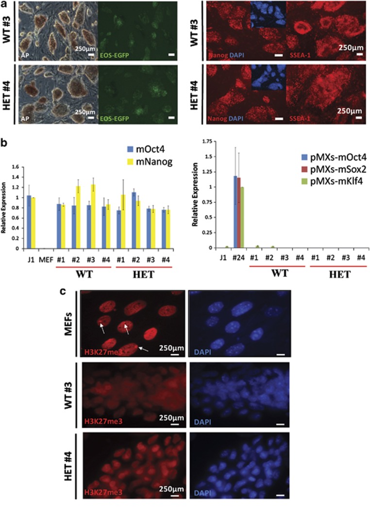
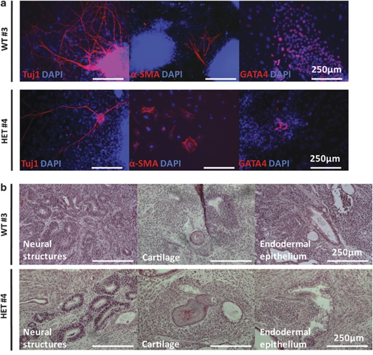
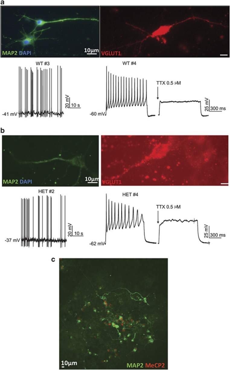
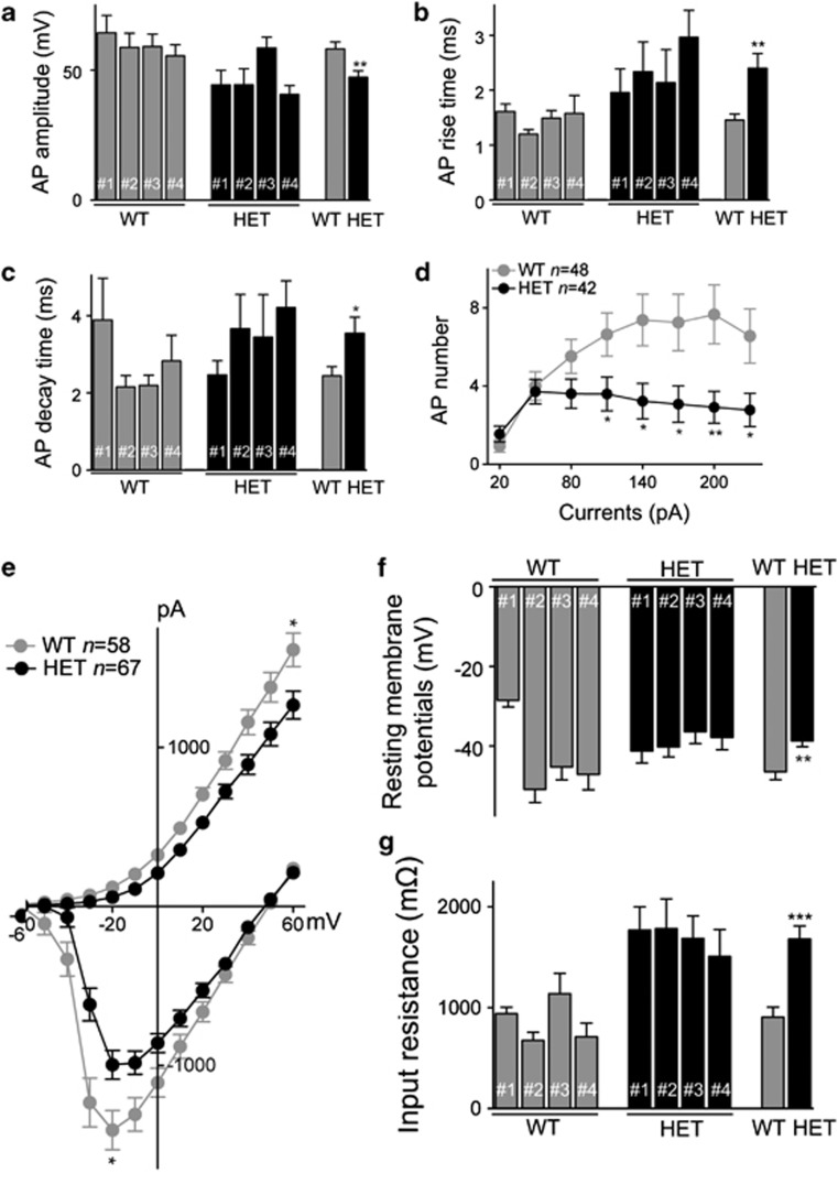
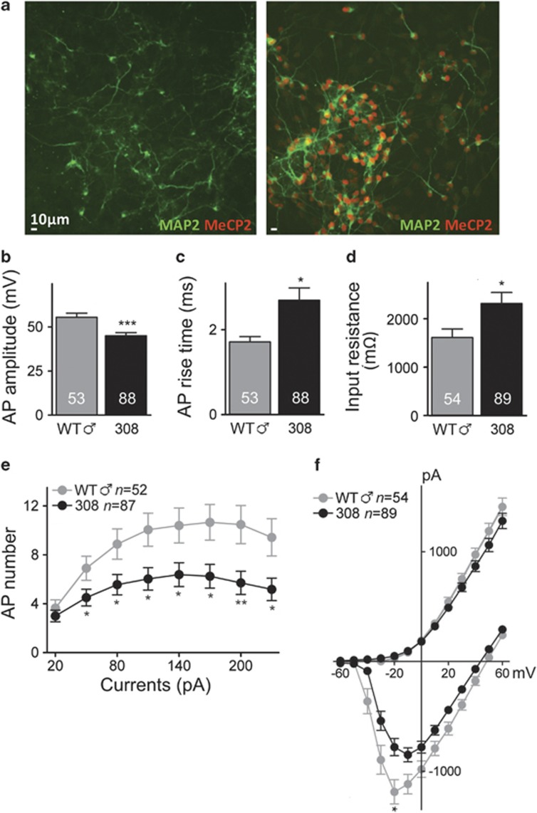
Rett syndrome (RTT) is a neurodevelopmental autism spectrum disorder caused by mutations in the methyl-CpG-binding protein 2 (MECP2) gene. Here, we describe the first characterization and neuronal differentiation of induced pluripotent stem (iPS) cells derived from Mecp2-deficient mice. Fully reprogrammed wild-type (WT) and heterozygous female iPS cells express endogenous pluripotency markers, reactivate the X-chromosome and differentiate into the three germ layers. We directed iPS cells to produce glutamatergic neurons, which generated action potentials and formed functional excitatory synapses. iPS cell-derived neurons from heterozygous Mecp2(308) mice showed defects in the generation of evoked action potentials and glutamatergic synaptic transmission, as previously reported in brain slices. Further, we examined electrophysiology features not yet studied with the RTT iPS cell system and discovered that MeCP2-deficient neurons fired fewer action potentials, and displayed decreased action potential amplitude, diminished peak inward currents and higher input resistance relative to WT iPS-derived neurons. Deficiencies in action potential firing and inward currents suggest that disturbed Na(+) channel function may contribute to the dysfunctional RTT neuronal network. These phenotypes were additionally confirmed in neurons derived from independent WT and hemizygous mutant iPS cell lines, indicating that these reproducible deficits are attributable to MeCP2 deficiency. Taken together, these results demonstrate that neuronally differentiated MeCP2-deficient iPS cells recapitulate deficits observed previously in primary neurons, and these identified phenotypes further illustrate the requirement of MeCP2 in neuronal development and/or in the maintenance of normal function. By validating the use of iPS cells to delineate mechanisms underlying RTT pathogenesis, we identify deficiencies that can be targeted for in vitro translational screens.


Figures

Figure 1
Figure 1
WT and Mecp2308 iPS cells are pluripotent. (a) WT #3 and HET #4 iPS cell lines express pluripotency markers alkaline phosphatase, Nanog and SSEA-1 by immunocytochemistry. (b) qRT-PCR analysis demonstrates that WT and HET iPS cell lines reactivate endogenous pluripotency loci (mouse mOct4 and mNanog) similar to the J1 ES cell line and also silence exogenous viral reprogramming transgenes (pMXs). The published partially reprogrammed EOS3F-24 (#24) iPS cell line is a positive control for transgene expression. Mouse embryonic fibroblasts are a differentiated cell type-negative control for pluripotency marker expression. (c) DAPI-colocalized H3K27me3 foci representing the inactive X-chromosome are absent in WT #3 and HET #4 iPS cells, in comparison with mouse embryonic fibroblasts (arrows). The H3K27me3 silencing mark is not present in all mouse embryonic fibroblasts because they are a mixture of male and female cells. Scale bars: 250 μm.
Figure 2
Figure 2
iPS cell lines are functionally pluripotent in vitro and in vivo. (a) WT #3 and HET #4 generate cells corresponding to the three germ layers (ectoderm, Tuj1/βIII-tubulin; mesoderm, α-SMA; and endoderm, GATA4) following EB-mediated in vitro differentiation. (b) WT #3 and HET #4 differentiate into the three germ layers (ectoderm, neural structures; mesoderm, cartilage; and endoderm, endodermal epithelium) in vivo in teratoma formation assays with immunodeficient mice. Scale bars: 250 μm.
Figure 3
Figure 3
iPS-derived cells display specific neuronal markers and generate TTX-sensitive action potentials, following differentiation directed to glutamatergic neurons. (a) WT iPS-derived cells express MAP2 and VGLUT1 as shown by immunocytochemistry (upper). A representative trace of a spontaneously discharging WT iPS-derived cell during current-clamp recording is shown in the lower left. Current-clamp recordings from a different WT iPS-derived cell are shown in the lower right pair of traces. In this otherwise silent cell, action potentials triggered by current injection (+110 pA) (left) were blocked by TTX (0.5 μM; right). (b) HET iPS-derived cells express MAP2 and VGLUT1 (upper). Representative traces during current-clamp recording show a spontaneously discharging HET iPS-derived cell (lower left) and another cell (lower right pair) in which action potentials, elicited by current injection (+110 pA), were blocked by TTX. (c) HET iPS-derived cultures show a mix of MeCP2+ and MeCP2– neurons (as indicated by staining for MAP2). Scale bars: 10 μm.
Figure 4
Figure 4
Action potential characteristics, voltage-activated currents, resting membrane potential and input resistance are altered in HET iPS-derived neurons compared with WT iPS-derived neurons. (a) Histogram showing average amplitude of action potentials in WT iPS-derived neurons (grey) and HET (black) iPS-derived neurons. The groups of bars on the left and middle show the four individual WT and HET sublines, respectively. Numbers of cells were 2, 11, 21 and 14 for WT #1,2,3,4, and 8, 9, 12 and 13 for HET #1,2,3,4, respectively. On right are the overall averages for WT (n=48 cells) and HET (n=42 cells). (b) Histogram of average rise time of action potentials in the WT and HET sublines and overall groups as in panel a. (c) Histogram of average decay time of action potentials in the WT and HET sublines and overall groups as in panel a. (d) A plot depicting the numbers of evoked action potentials elicited by depolarizing current steps from the overall groups of WT and HET sublines. The current steps were from +20 pA to +230 pA in +30-pA increments. (e) A plot showing average current–voltage relationships in WT compared with HET iPS-derived neurons. Recordings were made in voltage-clamp mode (holding potential −70 mV) and currents were elicited by a series of voltage steps from −60 to +60 mV. Early inward and late outward currents were measured as illustrated in the traces in Supplementary Figure 10. Maximum average inward currents and maximum outward currents of WT were compared with those of HET, *P<0.05. (f) Histogram of average resting membrane potentials in the WT and HET sublines and overall groups. (g) Histogram of average input resistance in the WT and HET sublines and overall groups. For resting membrane potential and input resistance, n=3, 15, 26 and 14 for WT #1,2,3,4, and n=13, 18, 18 and 18 for HET #1,2,3,4, respectively; totals WT (n=58) and HET (n=67). In this and all Figures, data points are mean ± s.e.m.; *P<0.05, **P<0.01 and ***P<0.001.
Figure 5
Figure 5
Hemizygous Mecp2308/y male iPS-derived neurons show deficits in action potential characteristics, excitability and voltage-activated Na+ currents. (a) Neuronally differentiated male ♂ WT (Mecp2+/y; WT ♂) and mutant (Mecp2308/y; 308) iPS-derived cells express MAP2. Immunofluorescence analysis using a C-terminal antibody unable to detect the C-terminal truncated MeCP2308 protein reveals that mutant iPS cell line 308 #1 is negative for MeCP2 expression (left). In contrast, WT ♂ #11 iPS-derived neurons express MeCP2 in all MAP2+ cells (right). Scale bars: 10 μm. (b) Histogram shows average action potential amplitude for total iPS-derived cell lines of WT ♂ (grey, n=53 neurons) and 308 (black, n=88 neurons). (c) Average action potential rise time from WT ♂ iPS-derived neurons and 308 iPS-derived neurons. (d) Average input resistances of WT ♂ and 308 iPS-derived neurons. (e) The plot shows average numbers of action potentials evoked by a series of depolarizing current steps from WT ♂ and 308 iPS-derived neurons. (f) A plot of average current–voltage relationships in WT ♂ compared with 308 iPS-derived neurons. *P<0.05, **P<0.01 and ***P<0.001.

References

    1. Amir RE, Van den Veyver IB, Wan M, Tran CQ, Francke U, Zoghbi HY. Rett syndrome is caused by mutations in X-linked MECP2, encoding methyl-CpG-binding protein 2. Nat Genet. 1999;23:185–188. - PubMed
    1. Hagberg B, Aicardi J, Dias K, Ramos O. A progressive syndrome of autism, dementia, ataxia, and loss of purposeful hand use in girls: Rett's syndrome: report of 35 cases. Ann Neurol. 1983;14:471–479. - PubMed
    1. Jung BP, Jugloff DG, Zhang G, Logan R, Brown S, Eubanks JH. The expression of methyl CpG binding factor MeCP2 correlates with cellular differentiation in the developing rat brain and in cultured cells. J Neurobiol. 2003;55:86–96. - PubMed
    1. Chen RZ, Akbarian S, Tudor M, Jaenisch R. Deficiency of methyl-CpG binding protein-2 in CNS neurons results in a Rett-like phenotype in mice. Nat Genet. 2001;27:327–331. - PubMed
    1. Calfa G, Percy AK, Pozzo-Miller L. Experimental models of Rett syndrome based on Mecp2 dysfunction. Exp Biol Med. 2011;236:3–19. - PMC - PubMed

Publication types

MeSH terms