. 2012 Jan 9;481(7382):469-74.
doi: 10.1038/nature10737.

X-ray structures of LeuT in substrate-free outward-open and apo inward-open states

Affiliations

X-ray structures of LeuT in substrate-free outward-open and apo inward-open states

Harini Krishnamurthy et al. Nature. .

Abstract

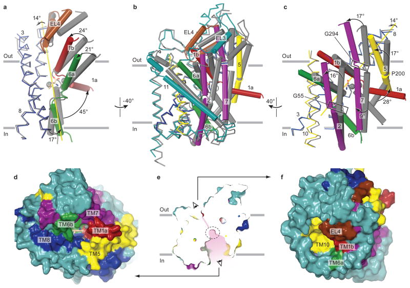
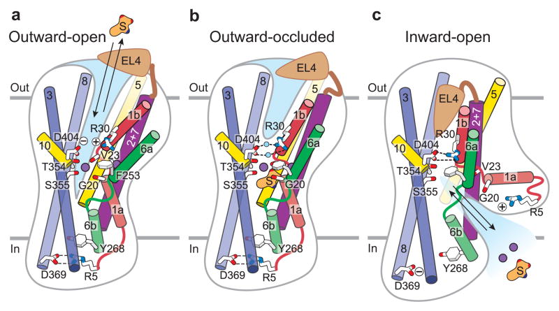
Neurotransmitter sodium symporters are integral membrane proteins that remove chemical transmitters from the synapse and terminate neurotransmission mediated by serotonin, dopamine, noradrenaline, glycine and GABA (γ-aminobutyric acid). Crystal structures of the bacterial homologue, LeuT, in substrate-bound outward-occluded and competitive inhibitor-bound outward-facing states have advanced our mechanistic understanding of neurotransmitter sodium symporters but have left fundamental questions unanswered. Here we report crystal structures of LeuT mutants in complexes with conformation-specific antibody fragments in the outward-open and inward-open states. In the absence of substrate but in the presence of sodium the transporter is outward-open, illustrating how the binding of substrate closes the extracellular gate through local conformational changes: hinge-bending movements of the extracellular halves of transmembrane domains 1, 2 and 6, together with translation of extracellular loop 4. The inward-open conformation, by contrast, involves large-scale conformational changes, including a reorientation of transmembrane domains 1, 2, 5, 6 and 7, a marked hinge bending of transmembrane domain 1a and occlusion of the extracellular vestibule by extracellular loop 4. These changes close the extracellular gate, open an intracellular vestibule, and largely disrupt the two sodium sites, thus providing a mechanism by which ions and substrate are released to the cytoplasm. The new structures establish a structural framework for the mechanism of neurotransmitter sodium symporters and their modulation by therapeutic and illicit substances.


Conflict of interest statement

Competing financial interests

The authors declare no competing financial interests.

Figures

Figure 1
Figure 1. Substrate-free, Na+-bound state is outward-open
a, Superposition of the outward-open and leucine-bound outward-occluded conformations. Outward-open structure is colored with Na+ ions as purple spheres. The outward-occluded structure with Na+ ions (spheres) is grey and leucine in stick representation. b, Schematic of scaffold and core domains, EL4, the pivot points of hinge movements in TMs 1 and 6 (solid black circles) and the substrate (S) and sodium sites (+). c, Superposition, as in panel a, illustrating that a ~9° rotation about an axis passing through the middle of the core domain (yellow arrow) describes the conformational change associated with opening to the outside. Pivot points are shown as in panel b. d, Rupture of extracellular gate interactions (grey dashed lines) in the outward-open structure. Two water molecules that bridge Arg 30 and Asp 404 in the outward-occluded state are shown as red spheres. e, Surface representation of the outward-open structure with the zig-zag pink line indicating a closed intracellular pathway. Leucine, where shown, is from the outward-occluded Leu-bound structure.
Figure 2
Figure 2. Sodium sites in outward-open state
a, The Na1 site showing positions of the coordinating residues within the framework of global changes in the outward-open structure. b, The Na2 site. Fo - Fc omit density is contoured at 4 σ and represented as green mesh. Coloring scheme and representations are the same as in Fig. 1. Dashed lines indicate interactions between sodium ions and coordinating atoms with distances in Å, solid black circles are approximate pivot points for hinge movement of helices and red and green cylinders define the TM1 and 6 helix axes, respectively.
Figure 3
Figure 3. Inward-open conformation
a–c, Superposition of inward-open and outward-occluded state structures using the scaffold domain. The overall changes shown in b are divided in two parts, a and c, for clarity. The extent of rotation for the key TMs between the outward-occluded and inward-open conformations are indicated in a and c. The axis of rotation of core domain with the exclusion of TM1a is depicted in yellow. d, Surface representation of the inward-open structure looking ‘up’ into the binding site from intracellular side as indicated in e. f, Surface representation of the inward-open structure showing the elements forming the extracellular thick gate. Color coding and representations are as in Fig. 1. Leucine and sodium, where shown, are from the outward-occluded Leu-bound structure.
Figure 4
Figure 4. Changes in gate, substrate and ion site interactions and coupling to helix movements
a, Comparison of the extracellular gating interactions in the inward-open and outward-occluded structures. View is from extracellular side. Polar contacts in the inward-open structure are shown as black dashed lines. b, Overall view of inward-open structure showing closed extracellular gate (box with solid lines) and open intracellular gate (box with dashed lines). c, Comparison of the intracellular gating interactions in the inward-open and outward-occluded structures. Interactions forming the intracellular gate in the outward-occluded state are shown as grey dashed lines. d, Changes in the central substrate binding site. In comparing the outward-occluded and inward-open structures, A22, T254, and F253 move away from the binding site. Distances were measured relative to leucine from the outward-occluded structure. e, A cartoon representing changes in the core domain relative to scaffold domain and location of hinges relative to position of substrate and ion binding sites is shown, with the pink lines indicating a closed extracellular pathway. f, Changes in the Na1 site. Distances of the coordinating residues from sodium ion of the outward-occluded structure are shown. g, Superposition of the inward-open and outward-occluded structures using TM7. TM1a of only the inward-open structure is shown for clarity. Coloring scheme as representations are as in Figure 1. Location of TM1 and TM6 hinges are shown as black spheres in c, d, e, and f.
Figure 5
Figure 5. Schematic of transport in LeuT
Shown are structural elements and gating residues instrumental to conformational changes associated with the transition from the outward-open (a) to the outward-occluded state (b) and the inward-open state (c). At present there is no crystal structure for an inward-occluded state and thus no schematic is provided.

References

    1. Hertting G, Axelrod J. Fate of tritiated noradrenaline at the sympathetic nerve-endings. Nature. 1961;192:172–173. - PubMed
    1. Nelson N. The family of Na+/Cl- neurotransmitter transporters. J Neurochem. 1998;71:1785–1803. - PubMed
    1. Saier MHJ. A functional-phylogenetic classification system for transmembrane solute transporters. Microbiol Mol Biol Rev. 2000;64:354–411. - PMC - PubMed
    1. Hahn MK, Blakely RD. Monoamine transporter gene structure and polymorphisms in relation to psychiatric and other complex disorders. Pharmacogenomics J. 2002;2:217–235. - PubMed
    1. Klimek V, et al. Reduced levels of norepinephrine transporters in the locus coeruleus in major depression. J Neurosci. 1997;17:8451–8458. - PMC - PubMed

Publication types

MeSH terms