. 2012 Jan 8;8(2):205-10.
doi: 10.1038/nchembio.764.

Timing facilitated site transfer of an enzyme on DNA

Affiliations

Timing facilitated site transfer of an enzyme on DNA

Joseph D Schonhoft et al. Nat Chem Biol. .

Abstract

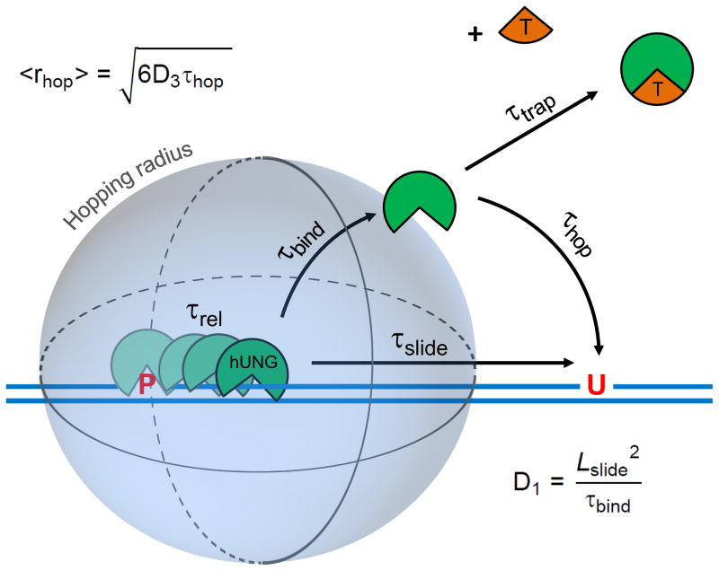
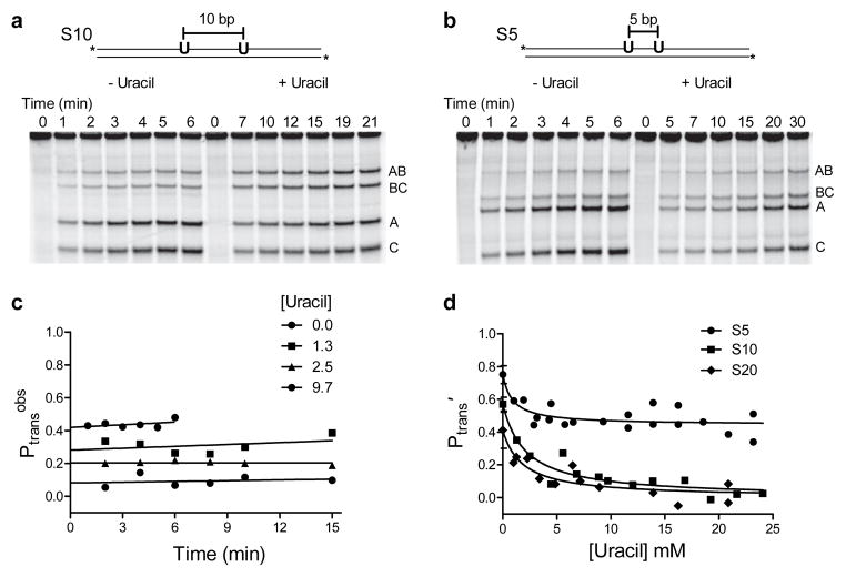
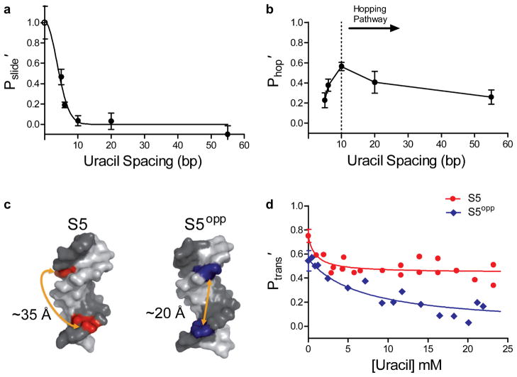
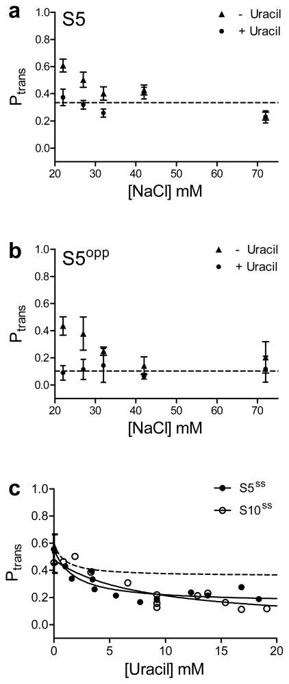
Many enzymes that react with specific sites in DNA have the property of facilitated diffusion, in which the DNA chain is used as a conduit to accelerate site location. Despite the importance of such mechanisms in gene regulation and DNA repair, there have been few viable approaches to elucidate the microscopic process of facilitated diffusion. Here we describe a new method in which a small-molecule trap (uracil) is used to clock a DNA repair enzyme as it hops and slides between damaged sites in DNA. The 'molecular clock' provides unprecedented information: the mean length for DNA sliding, the one-dimensional diffusion constant, the maximum hopping radius and the time frame for DNA hopping events. In addition, the data establish that the DNA phosphate backbone is a sufficient requirement for DNA sliding.


Conflict of interest statement

Competing financial interests

The authors declare no competing financial interests.

Figures

Figure 1
Figure 1
The molecular clock approach for timing pathways for facilitated diffusion on DNA. The molecular clock begins when hUNG is released from the abasic site product (P) produced from its excision of uracil from one site in DNA and then begins its journey to the second site (U) by either hopping or sliding. The rate-limiting product release step occurs with a characteristic release time (τrel), after which hUNG is bound non-specifically to DNA with a lifetime τbind = 1/koff. The hopping pathway involves one or more dissociation events from non-specific DNA to generate a free enzyme molecule that is still very close to the original DNA chain from which it dissociated. The free lifetime (τhop) of a dissociated enzyme molecule following the hopping pathway will depend on its 3D diffusion constant (D3) and its distance from the DNA chain (r). In contrast, the sliding pathway involves direct transfer of an enzyme molecule to the second site without dissociation. The sliding length (Lslide) will depend on how long the enzyme remains bound to non-specific DNA (τbind) and its 1D diffusion constant (D1). The timing mechanism of the clock is provided by the concentration and diffusion constant of a small molecule trap (T) that can capture hopping, but not sliding enzyme molecules. The molecular clock can be used to calculate D1 and the hopping radius (<rhop>), which is the distance at which half of the hopping enzyme molecules are trapped and half find the second target site successfully by hopping (see text).
Figure 2
Figure 2
Facilitated site transfer by human uracil DNA glycosylase (hUNG). In all facilitated transfer assays the hUNG concentration is 5–20 pM and the DNA substrate concentration is 40 nM. (a) Facilitated site transfer of hUNG between two uracil sites on the same DNA strand separated by 10 bps (S10). Reactions in the absence and presence of 10 mM uracil are shown. Facilitated transfer is qualitatively indicated by an excess of double excision fragments (A and C) relative single site excision products (AB and BC). (b) Facilitated site transfer by hUNG between sites separated by 5 bp (S5) in the absence and presence of 10 mM uracil. (c) Observed probability of site transfer (Ptransobs) as a function of time and uracil concentration for the substrate with a 10 bp site separation. Linear extrapolation to the y axis provides the true transfer probability at zero time (Ptrans). (d) Ptrans′ as a function of increasing uracil for substrates with 5, 10 and 20 bp site spacings. Each data point represents an individual experiment as in panels a and b and the prime notation indicates correction for the efficiency of excision (see text). The non-linear least squares fits in (d) use a kinetic partitioning model that relates the dependence of the total transfer probability (Ptrans′ = Pslide′ + Phop′) to the uracil trap concentration (Supplementary Methods). Uncut gel images are shown in Supplementary Fig. 6. Error bars represent the mean ± 1 s.d. of at least three independent trials.
Figure 3
Figure 3
Probability of sliding (Pslide′) and hopping (Phop′) as a function of site spacing and strand positioning of uracils. The individual contributions of the sliding and hopping pathways at each site spacing were determined by measuring the limiting Ptrans′ at high concentrations of uracil (sliding only), and the difference between the Ptrans at zero and high concentrations of uracil (hopping only). (a) Site spacing dependence of Pslide′. The non-linear least squares fit to the function Pslide′ = SN is shown, where S is a constant and N is the site spacing in bp squared. The maximal efficiency of a sliding enzyme at zero spacing is 1.0 and is equivalent to the efficiency of excision (open circle). The mean sliding length (Lslide) was calculated from the spacing at Pslide′ = 0.5 and was determined to be 4.2 ± 0.8 bp where the error represents the 95% confidence interval of the least squares fit. (b) Site spacing dependence of Phop′. The dotted line indicates the approximate base pair separation where a change from a predominantly hopping to sliding pathway for facilitated transfer occurs. (c) Structural models of B-DNA helices illustrating the position and approximate distances of two uracils positioned on the same (S5) or opposite DNA strand (S5opp). Images were made in Pymol using PDB I.D. 2L8Q. (d) Transfer efficiencies for S5 and S5opp as a function of uracil concentration. Error bars represent the mean ± 1 s.d. of at least three independent trials.
Figure 4
Figure 4
Site transfer dependence with increasing salt and in the context of single stranded DNA. No correction for the cleavage efficiency (E) was made in these transfer measurements. (a & b) Transfer probabilities for substrate S5 and S5opp with and without the addition of 10 mM uracil as a function of increasing [NaCl]. For both substrates with increasing NaCl, the transfer probabilities plateau at the level observed in the presence of uracil. The average salt-independent values for Ptrans in the presence of uracil are indicated by the dotted lines in panels (a) & (b). For both (a) & (b) in the presence of uracil, the Ptrans value at 22 mM NaCl is the plateau average with increasing uracil (Fig. 2d and Fig. 4b). (c) hUNG slides on single stranded DNA. Transfer probabilities were measured for a 90mer substrate with uracils positioned 5 and 10 bp apart (S5ss, S10ss). The plateau in Ptrans for S5ss andS10ss equals 0.22 ± 0.04 and 0.16 ± 0.05 respectively. For comparison, the dashed line is the theoretical fit to the data for the duplex form S5 (also with no correction for the cleavage efficiency). Error bars represent the mean ± 1 s.d. of at least three independent trials.

References

    1. Riggs AD, Bourgeois S, Cohn M. The lac represser-operator interaction: III. Kinetic studies. J Mol Biol. 1970;53:401–417. - PubMed
    1. Berg OG, Winter RB, Von Hippel PH. Diffusion-driven mechanisms of protein translocation on nucleic acids. 1 Models and theory. Biochemistry. 1981;20:6929–6948. - PubMed
    1. Gowers DM, Wilson GG, Halford SE. Measurement of the contributions of 1D and 3D pathways to the translocation of a protein along DNA. Proc Natl Acad Sci U S A. 2005;102:15883–15888. - PMC - PubMed
    1. Stanford NP, Szczelkun MD, Marko JF, Halford SE. One- and three-dimensional pathways for proteins to reach specific DNA sites. EMBO J. 2000;19:6546–6557. - PMC - PubMed
    1. Hedglin M, O’Brien PJ. Human Alkyladenine DNA Glycosylase Employs a Processive Search for DNA Damage. Biochemistry. 2008;47:11434–11445. - PMC - PubMed

Publication types

Associated data