Case Reports
. 2012 Jan 16;209(1):29-34.
doi: 10.1084/jem.20110896. Epub 2012 Jan 9.

A novel primary human immunodeficiency due to deficiency in the WASP-interacting protein WIP

Affiliations
Case Reports

A novel primary human immunodeficiency due to deficiency in the WASP-interacting protein WIP

Gaetana Lanzi et al. J Exp Med. .

Abstract

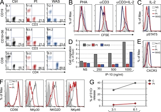
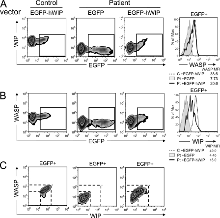
A female offspring of consanguineous parents, showed features of Wiskott-Aldrich syndrome (WAS), including recurrent infections, eczema, thrombocytopenia, defective T cell proliferation and chemotaxis, and impaired natural killer cell function. Cells from this patient had undetectable WAS protein (WASP), but normal WAS sequence and messenger RNA levels. WASP interacting protein (WIP), which stabilizes WASP, was also undetectable. A homozygous c.1301C>G stop codon mutation was found in the WIPF1 gene, which encodes WIP. Introduction of WIP into the patient's T cells restored WASP expression. These findings indicate that WIP deficiency should be suspected in patients with features of WAS in whom WAS sequence and mRNA levels are normal.


Figures

Figure 1.
Figure 1.
Functional characterization of WIP deficiency. (A) Evaluation of the proportion of T, B, and NK cells on gated lymphocytes in whole blood (top and middle row), and of CD4- and CD8-expressing cells on the CD3+ gated cells (bottom row) of a control (Ctrl), the patient (Pt), and a WAS-null patient. Data are representative of three independent analyses. (B) T cell proliferation to PHA and anti-hCD3 with or without rIL-2, as assessed by CFSE dilution. PBMCs were used and FACS analysis was performed on CD3+ gated cells. The solid gray histogram represents exemplificative profile of unstimulated cells. An overlapping profile was obtained from unstimulated cells of control and WAS patients (not depicted). The red, gray, and blue profiles represent CFSE content in stimulated cells from patient, control, and a WAS-null patient, respectively. Data are representative of two independent experiments. (C) PBMCs were stimulated with IL-2, and gated CD3+ cells were analyzed by FACS for intracellular pSTAT5 using anti-pY694STAT5. The solid line represents the signal in unstimulated patient’s cells, which was comparable to that of unstimulated control and WAS-null patient cells (not depicted). The pSTAT5 signal in the rIL-2 stimulated cells from patient, control, and WAS patient are indicated in red, gray, and blue, respectively. Data are representative of three independent experiments. (D) Migration of PHA blasts from a healthy control, the patient and from a WAS-null patient toward the chemokine IP-10. The gray, red, and blue histograms represent migration of cells from control, patient, and WAS-null patient, respectively. Data are representative of two independent experiments. (E) Expression of the IP-10 receptor CXCR3 on PHA blasts from patient, control, and WAS-null patient. Isotype control staining of patient PHA blasts is shown by the filled histogram and was comparable to that of control T cells and WAS patient (not depicted). The CXCR3 signals in cells from patient, control, and WAS patient are indicated in red, gray, and blue, respectively. Data are representative of two independent experiments. (F) Expression of CD56, NKp30, NKGD2, and NKp46 on NK cells from patient (red) and control (gray). The filled histogram represents isotype control staining of patient NK cells, and was comparable to that of control NK cells (not depicted). Data are representative of two analyses. (G) Cytolytic activity by patient and control NK cell lines against the LCL 721.221 target cells, measured as the percentage of net 51Cr release at effector (E) to target (T) ratios of 3:1 and 6:1. Data are representative of two independent experiments on the same expanded NK cells.
Figure 2.
Figure 2.
WIP is mutated in the patient. (A and C) Western blot analysis of WASP (A) and WIP (C) in lysates of PHA T cell blasts. Numbers at the bottom indicate densitometric quantification corrected for the actin signal. Data are representative of two experiments. (B and D) qPCR analysis of mRNA expression of WAS (B) and WIPF1 (D) in PHA T blasts, relative to the housekeeping gene GAPDH, normalized to 1 for the mean of 25 controls. The gray area indicates the range for the 25 controls. Data are representative of three experiments conducted on triplicates. (E) Genomic organization of WIPF1 (top) and protein structure (bottom) of WIP showing the localization of the mutation and the resulting amino acidic change (in red) from Serine to a premature termination codon (S434X). The genomic organization shows coding exons, indicated with Roman numbers, and 5′ and 3′ untranslated regions (UTR). Protein structure includes known WIP-interacting protein domains and WASP homology 2 (WH2). (F) Electropherogram depicting the homozygous c.1301C>G mutation in exon 6 in the patient and the presence of the same mutation in the heterozygous state in the mother.
Figure 3.
Figure 3.
Introduction of WIP restores WASP levels in the patient T cells. (A and B) Two-color FACS analysis of expression of WIP (A) and WASP (B) versus EGFP in PHA T cell blasts transfected with vectors encoding EGFP-hWIP for patient and control and with EGFP alone for the patient. Histograms depicting WIP and WASP expression in gated EGFP+ cells are shown in the right panels. (C) Two-color FACS analysis of WASP versus WIP expression in gated EGFP+ cells. Data are representative of two different analyses.

Similar articles

Cited by

References

    1. Antón I.M., de la Fuente M.A., Sims T.N., Freeman S., Ramesh N., Hartwig J.H., Dustin M.L., Geha R.S. 2002. WIP deficiency reveals a differential role for WIP and the actin cytoskeleton in T and B cell activation. Immunity. 16:193–204 10.1016/S1074-7613(02)00268-6 - DOI - PubMed
    1. Castriconi R., Dondero A., Cilli M., Ognio E., Pezzolo A., De Giovanni B., Gambini C., Pistoia V., Moretta L., Moretta A., Corrias M.V. 2007. Human NK cell infusions prolong survival of metastatic human neuroblastoma-bearing NOD/scid mice. Cancer Immunol. Immunother. 56:1733–1742 10.1007/s00262-007-0317-0 - DOI - PMC - PubMed
    1. Curcio C., Pannellini T., Lanzardo S., Forni G., Musiani P., Antón I.M. 2007. WIP null mice display a progressive immunological disorder that resembles Wiskott-Aldrich syndrome. J. Pathol. 211:67–75 10.1002/path.2088 - DOI - PubMed
    1. de la Fuente M.A., Sasahara Y., Calamito M., Antón I.M., Elkhal A., Gallego M.D., Suresh K., Siminovitch K., Ochs H.D., Anderson K.C., et al. 2007. WIP is a chaperone for Wiskott-Aldrich syndrome protein (WASP). Proc. Natl. Acad. Sci. USA. 104:926–931 10.1073/pnas.0610275104 - DOI - PMC - PubMed
    1. Gallego M.D., de la Fuente M.A., Anton I.M., Snapper S., Fuhlbrigge R., Geha R.S. 2006. WIP and WASP play complementary roles in T cell homing and chemotaxis to SDF-1alpha. Int. Immunol. 18:221–232 10.1093/intimm/dxh310 - DOI - PubMed

Publication types

MeSH terms

Substances