. 2012 Jan 8;19(2):158-63.
doi: 10.1038/nsmb.2208.

Structure of a KirBac potassium channel with an open bundle crossing indicates a mechanism of channel gating

Affiliations

Structure of a KirBac potassium channel with an open bundle crossing indicates a mechanism of channel gating

Vassiliy N Bavro et al. Nat Struct Mol Biol. .

Abstract

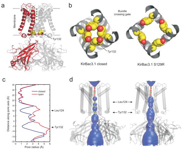
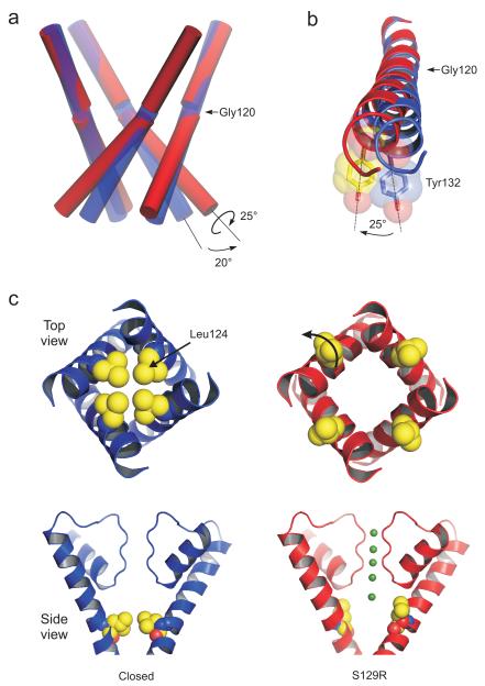
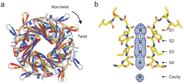
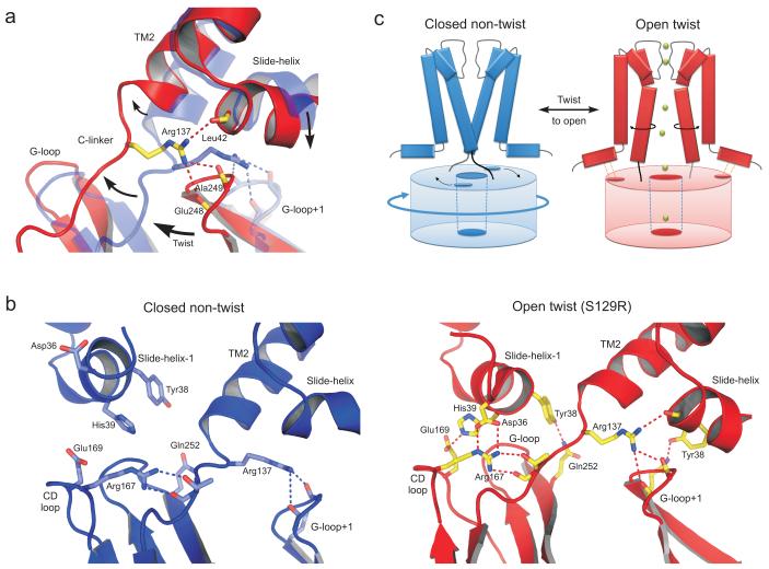
KirBac channels are prokaryotic homologs of mammalian inwardly rectifying (Kir) potassium channels, and recent crystal structures of both Kir and KirBac channels have provided major insight into their unique structural architecture. However, all of the available structures are closed at the helix bundle crossing, and therefore the structural mechanisms that control opening of their primary activation gate remain unknown. In this study, we engineered the inner pore-lining helix (TM2) of KirBac3.1 to trap the bundle crossing in an apparently open conformation and determined the crystal structure of this mutant channel to 3.05 Å resolution. Contrary to previous speculation, this new structure suggests a mechanistic model in which rotational 'twist' of the cytoplasmic domain is coupled to opening of the bundle-crossing gate through a network of inter- and intrasubunit interactions that involve the TM2 C-linker, slide helix, G-loop and the CD loop.


Figures

Figure 1
Figure 1
Structure of the KirBac3.1 S129R mutant in an apparently open conformation. (a) Overall structure of S129R mutant with one subunit highlighted in red for clarity. Ions within the selectivity filter are shown in green and residue Tyr132 which forms the primary gate at the bundle-crossing is shown as CPK spheres in all four subunits. (b) Bottom up view of the bundle-crossing gate in an example of a closed state KirBac3.1 (PDB 2WLJ) and the S129R mutant channel. Tyr132 side chains are shown as CPK spheres. (c) Pore radius profile of a closed state KirBac3.1 (PDB 2WLJ) (Closed, blue) and the S129R mutant where the Arg129 side chain has been replaced by a serine (Open, red). (d) The pore-lining surface and structure of the open and closed KirBac3.1 channels with the position of the major constrictions Tyr132 and Leu124 marked by arrows.
Figure 2
Figure 2
Bending of the inner TM2 helices during channel opening (a) Conformational changes of the inner helices from the closed state (blue cylinders) to the conformation (red cylinders) seen in the S129R mutant. The lower section of TM2 kinks by up to 20° at the conserved glycine hinge (Gly120) and also rotates around its helical axis by 25° when viewed from below. (b) The clockwise rotation of TM2 is shown viewed from below. Overlay of TM2 helices from the S129R structure (red) and a closed state Kir3.1 (blue, PDB 2WLJ). Tyr132 is shown as a transparent CPK sphere. (c) Opening of the inner cavity constriction formed by Leu124. Top and side views are shown on the S129R structure (red) and a closed state KirBac3.1 (blue, PDB 2WLJ). The position of ions within the filter of the S129R structure is shown in green. The rotation of TM2 moves Leu124 away from the pore. Leu124 is equivalent to the rectification control site in TM2 of eukaryotic Kir channels.
Figure 3
Figure 3
S129R is in a twisted yet conductive conformation. (a) The intracellular assemblies of a non-twist (blue, PDB 2WLJ) and twist (yellow, PDB 2X6C) conformation are viewed from the top relative to their superimposed pore domains (not shown). The relative position of the S129R CTD is also overlaid (red) showing that it is in the ‘twist’ conformation. (b) Fo-Fc OMIT map of electron density in the selectivity filter of the S129R mutant channels contoured at ~3σ showing clear density in all four binding sites, as well as the cavity site.
Figure 4
Figure 4
Interaction network between the CTD and the TMD. (a) The C-linker in the twist S129R structure (red) is displaced by up to 5Å compared to the closed non-twist structure (PDB 2WLJ, transparent blue). During twisting and opening Arg137 shifts its interaction with backbone carbonyls on the G-loop of the adjacent subunit (G-loop+1) to include an intrasubunit interaction with the slide-helix. The major motions are indicated by arrows. (b) Comparison of the network of interactions formed in the S129R twist open conformation (red) with those in a closed non-twist conformation (PDB 2WLJ). Movement of the slide helix and displacement of the C-linker allow an extensive network of inter and intra-subunit interactions to form (see also Supplementary Movie 2) (c) Cartoon of gating model proposed by the motions observed in the S129R structure. The twist conformation of the CTD pre-tensions the C-linker and also brings the slide-helix into register with the CD-loop (dot shown on CTD), thereby coupling rotational movement of the CTD to opening at the bundle-crossing gate.

References

    1. Hibino H, et al. Inwardly rectifying potassium channels: their structure, function, and physiological roles. Physiol Rev. 2010;90:291–366. - PubMed
    1. Bichet D, Haass FA, Jan LY. Merging functional studies with structures of inward-rectifier K+ channels. Nat Rev Neurosci. 2003;4:957–67. - PubMed
    1. Swartz KJ. Towards a structural view of gating in potassium channels. Nat Rev Neurosci. 2004;5:905–16. - PubMed
    1. Kuo A, et al. Crystal structure of the potassium channel KirBac1.1 in the closed state. Science. 2003;300:1922–6. - PubMed
    1. Kuo A, Domene C, Johnson LN, Doyle DA, Venien-Bryan C. Two different conformational states of the KirBac3.1 potassium channel revealed by electron crystallography. Structure. 2005;13:1463–72. - PubMed

Additional references for methods section

    1. Collaborative Computational Project, Number 4 The CCP4 Suite: Programs for Protein Crystallography. Acta Cryst D. 1994;50:760–763. - PubMed
    1. Evans PR. Scaling and assessment of data quality. Acta Cryst D. 2006;62:82–82. - PubMed
    1. McCoy AJ, et al. Phaser crystallographic software. J Appl Cryst. 2007;40:658–674. - PMC - PubMed
    1. Emsley P, Lohkamp B, Scott WG, Cowtan K. Features and development of Coot. Acta Cryst D. 2010;66:486–501. - PMC - PubMed
    1. Blanc E, et al. Refinement of severely incomplete structures with maximum likelihood in BUSTER-TNT. Acta Cryst D. 2004;60:2210–2221. - PubMed

Publication types

MeSH terms

Associated data

LinkOut - more resources