. 2012 Sep 20;31(38):4245-54.
doi: 10.1038/onc.2011.586. Epub 2012 Jan 9.

Chk1 phosphorylation of Metnase enhances DNA repair but inhibits replication fork restart

Affiliations

Chk1 phosphorylation of Metnase enhances DNA repair but inhibits replication fork restart

R Hromas et al. Oncogene. .

Erratum in

  • Oncogene. 2014 Jan 23;33(4):536. Laitao, A [corrected to Leitao, A]

Abstract

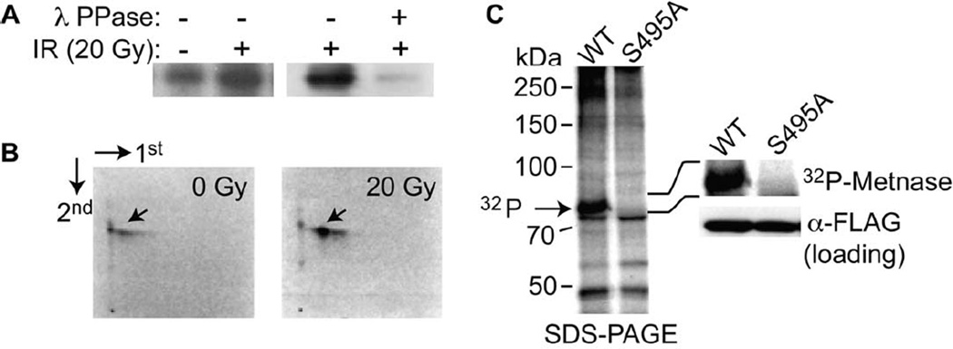
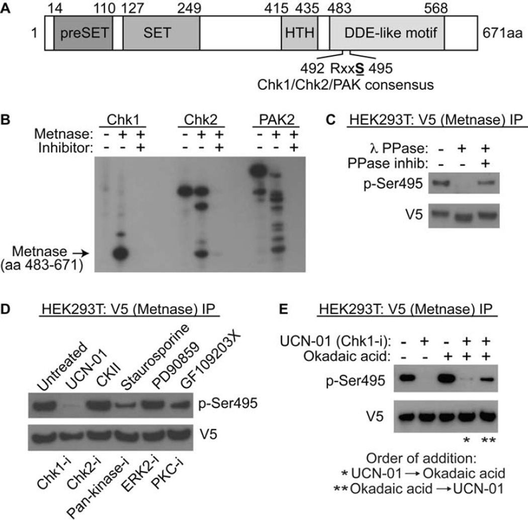
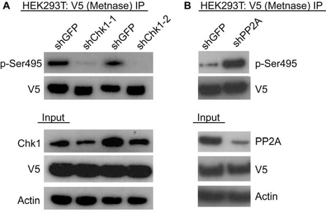
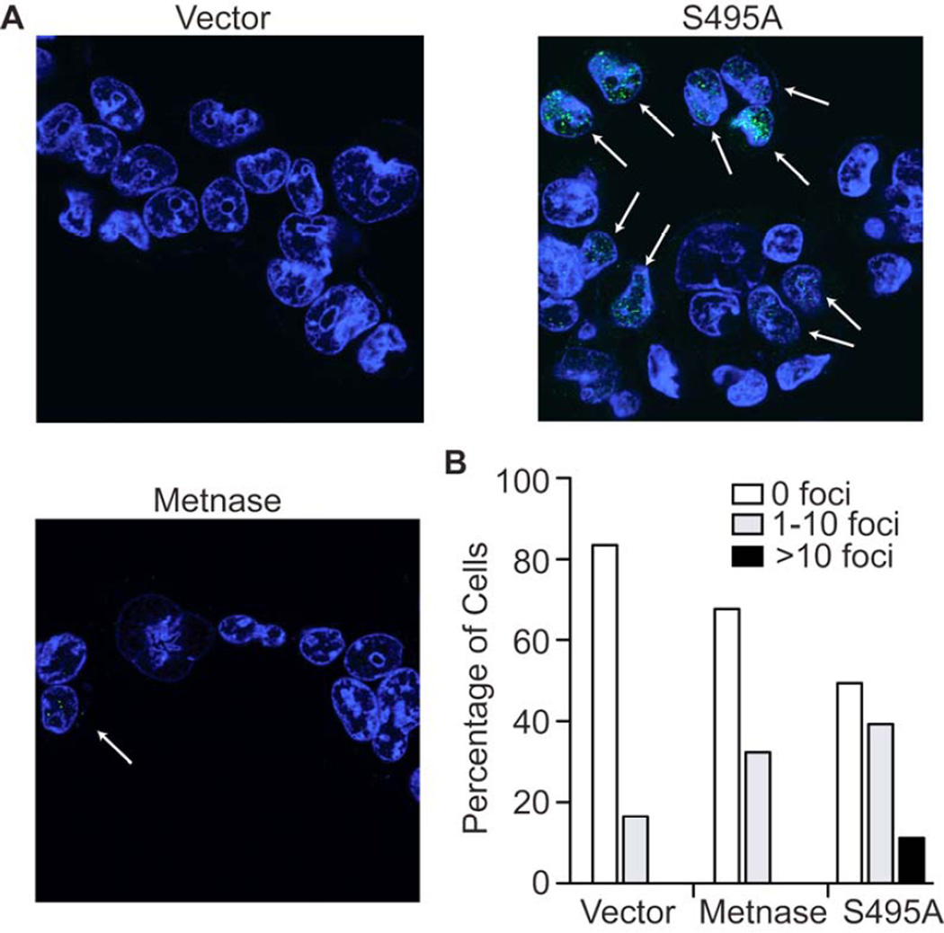
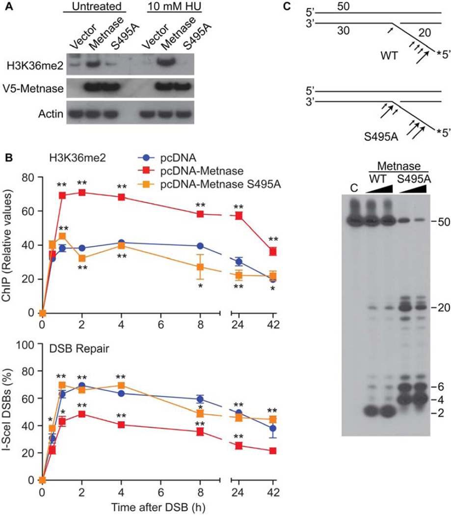
Chk1 both arrests replication forks and enhances repair of DNA damage by phosphorylating downstream effectors. Although there has been a concerted effort to identify effectors of Chk1 activity, underlying mechanisms of effector action are still being identified. Metnase (also called SETMAR) is a SET and transposase domain protein that promotes both DNA double-strand break (DSB) repair and restart of stalled replication forks. In this study, we show that Metnase is phosphorylated only on Ser495 (S495) in vivo in response to DNA damage by ionizing radiation. Chk1 is the major mediator of this phosphorylation event. We had previously shown that wild-type (wt) Metnase associates with chromatin near DSBs and methylates histone H3 Lys36. Here we show that a Ser495Ala (S495A) Metnase mutant, which is not phosphorylated by Chk1, is defective in DSB-induced chromatin association. The S495A mutant also fails to enhance repair of an induced DSB when compared with wt Metnase. Interestingly, the S495A mutant demonstrated increased restart of stalled replication forks compared with wt Metnase. Thus, phosphorylation of Metnase S495 differentiates between these two functions, enhancing DSB repair and repressing replication fork restart. In summary, these data lend insight into the mechanism by which Chk1 enhances repair of DNA damage while at the same time repressing stalled replication fork restart.


Figures

Figure 1
Figure 1. IR-induced phosphorylation of Metnase
A. Cells over-expressing Flag-Metnase were incubated 32P-orthophosphoric acid for 1 hr, and then exposed to 20 gy ionizing radiation (IR). Metnase was immunoprecipitated using anti-Flag, electorphoretically separated, and autoradiographed. The loss of signal after λ-phosphatase implies a direct phosphorylation event. B. Two dimensional phosphopeptide mapping analysis of 32P-Metnase following IR treatment demonstrated only one species, and this species was induced following IR. This phosphopeptide was ioslated and analyzed for amino acid sequence (Supplemental Fig. 1). The phosphorylation occurred at S495. C. Phosphorylation analysis of Metnase and the S495A mutant in vivo after 20 gy IR. Wild-type (wt) and the S495A mutant of Metnase were immunoprecipitated after 32P labeling. Only the wt Metnase was phosphophorylated, shown in magnified form on the right of the panel.
Figure 2
Figure 2. Phosphorylation of Metnase by Chk1 in vitro
A. Schematic diagram of Metnase. The PreSET domain contains a cysteine- and histidine-rich putative Zn++ binding motif, and the SET domain has the histone methylase motif. The transposase nuclease domain contains the two conserved motifs, HTH and DDE-like (in Metnase this is DDN, Roman et al 2008). The S495 is within RxxS Chk1/2/Pak consensus sites. B. Chk1 phosphorylates Metnase in vitro much more efficiently than Chk2 and PAK2. In vitro phosphorylation of pure recombinant Metnase by pure recombinant Chk1, Chk2 and PAK2 kinases, with and without their inhibitors (UCN-01, CKII, and staurosporin, respectively), is shown. C. The anti-phosho-Ser495 (anti-pS495) antibody recognizes the phosphorylation of Metnase. V5-tagged wt Metnase was immunoprecipitated from human 293T cells using the V5 tag, and treated with 400U λ-protein phosphatase in the presence or absence of phosphatase inhibitors (as described in Methods). The immunoprecipitate was analyzed for the presence of phosphorylated S495 using anti-sera specific for the phoshorylated S495. The loss of signal in the presence of phosphatasae implies the direct phosphorylation of Metnase at S495. D. Metnase phosphorylation in vivo in the presence of kinase inhibitors, listed above the panel. Below the panel are the types of kinases inhibited. The Chk1 and to a lesser extent the pan-kinase inhibitor block the phosphporylation of S495 on Metnase as detected by western analysis using anti-pSer495 after immunoprecipitation of V5-tagged wt Metnase. The samples are as follows: 1- no treatment; 2- UCN-01 (Chk1 inhibitor); 3- CKII (Chk2 inhibitor); staurosporine (pan-kinase inhibitor, including PAK2 and Chk1); PD90859 (ERK2 inhibitor); GF109203X (PKC inhibitor). E. The PP2A inhibitor Okadaic acid reverses the ability of the Chk1 kinase inhibitor UCN-01 to inhibit phosphorylation of S495 on Metnase. After immunoprecipitation of V5-tagged wt Metnase from cells pre-treated with UCN-01, okadic acid, or both, the pS495 was detected using western analysis. Okadaic acid was effective only when it was added prior to UCN-01.
Figure 3
Figure 3. Cellular phosphorylation of Metnase by Chk1, and dephosphorylation by PP2A
Repressing expression of Chk1 blocks Metnase S495 phosphorylation. V5-tagged Metnase was immunoprecipitated from cells with and without repression of Chk1 using specific siRNAs. The presence of phosphorylated Metnase was detected using western analysis with the anti-pS495. B. Repressing expression of PP2A enhances Metnase S495 phosphorylation. V5-tagged Metnase was immunoprecipitated from cells with and without siRNA repression of PP2A. The presence of phosphorylated Metnase was detected using western analysis with the anti-pS495.
Figure 4
Figure 4. Phosphorylation of Metnase S495 promotes its association with chromatin after IR but not after replication stress
A. Western analysis of chromatin-associated wild-type and S495A Metnase. Cells expressing FLAG-tagged Metnase or the S495A mutant were treated with 10 Gy IR, and chromatin was isolated at indicated times. Ku70 and histone H3 were used as nuclear protein loading controls. Expression of wild-type and S495A Metnase, analyzed by Western blot in total cell extracts, is shown to the right. B. V5-tagged wild-type or S495A Metnase were immunoprecipitated with anti-V5 and phospho-S495 Metnase was detected by Western blot (top panel). Middle and lower panels are V5-Metnase input and actin loading controls, respectively. C. Cells expressing FLAG-tagged Metnase were treated with 10 Gy IR, 10 mM HU, or mock treated, and phospho-S495 Metnase in chromatin-bound or unbound fractions was detected by Western blot. D. Chromatin association of wild-type and S495A Metnase as measured by coimmunoprecipitation analysis. FLAG-tagged wild-type or S495A Metnase were immunoprecipitated with anti-FLAG (middle panel) and their co-immunoprecipitation with histone H3 (top panel) was detected by Western blot. The lower panel shows input Metnase levels prior to immunoprecipitation. E. ChIP analysis of Metnase recruitment to a single DSB induced by I-SceI nuclease in HT1904 cells transfected with empty vector or over-expressing wild-thype or S495A Metnase (Fnu et al, 2011). ChIP was quantified by real-time PCR at indicated times after DSB induction. * P <0.05, ** P <0.01.
Figure 5
Figure 5. Expression of the S495A Metnase mutant increases replication fork recovery
A. Confocal immunofluorescence of BRDU foci (green) in 293T cells expressing either wt or S495A Metnase species after recovery from exposure to hydroxyurea. The foci represent replication forks newly incorporating BRDU after hydroxyurea has been removed. B. Quantifying cells expressing BRDU foci demonstrates that cells with S495A Metnase species have significantly more foci than with wt Metnase. More than 500 cells were counted on at least 5 slides for each experimental arm.
Figure 6
Figure 6. Metnase phosphorylation regulates its biochemoical activities
Metnase dimethylation of histone 3 lysine 35 (H3k36) and subsequent enhancement of DSB repair requires S495 phosphorylation. A. Western analysis of H3K36 dimethyltion (me2) by wt and S495A Metnase after DNA damage induced by hydroxyurea (HU). B. ChIP analysis (upper panel) over time after induction of a single DSB by ISCE-I for the adjacent presence of H3K36me2 with over-expression of wt or S495A Metnase (Fnu et al, 2011). The lower panel shows real time PCR assessment of the re-ligation of this induced single DSB over time. * P <0.05, ** p<0.01. C. Flap cleavage nuclease activities of pure isolated eukaryotic wt and S495A Metnase. The upper schematic diagrams the altered S495A flap nuclease activity. The size of the arrows correlates with the amount of endonucleolysis.

Similar articles

Cited by

References

    1. Agrawal A, Eastman QM, Schatz DG. Transposition mediated by RAG1 and RAG2 and its implications for the evolution of the immune system. Nature. 1998;394:744–751. - PubMed
    1. Boulikas T. Nuclear localization signals (NLS) Crit Rev Eukaryot Gene Expr. 1993;3:193–227. - PubMed
    1. Arnold K, Kiefer F, Kopp J, Battey JN, Podvinec M, Westbrook JD, Berman HM, Bordoli L, Schwede T. The Protein Model Portal. J. Struct. Funct. Genomics. 2009;10:1–8. - PMC - PubMed
    1. Beck BD, Lee SS, Williamson E, Hromas RA, Lee SH. Biochemical characterization of metnase's endonuclease activity and its role in NHEJ repair. Biochemistry. 50:4360–4370. - PMC - PubMed
    1. Brzeska H, Knaus UG, Wang ZY, Bokoch GM, Korn ED. p21-activated kinase has substrate specificity similar to Acanthamoeba myosin I heavy chain kinase and activates Acanthamoeba myosin I. Proc Natl Acad Sci U S A. 1997;94:1092–1095. - PMC - PubMed

Publication types