. 2012 Jan 8;18(2):244-51.
doi: 10.1038/nm.2598.

AICAR prevents heat-induced sudden death in RyR1 mutant mice independent of AMPK activation

Affiliations

AICAR prevents heat-induced sudden death in RyR1 mutant mice independent of AMPK activation

Johanna T Lanner et al. Nat Med. .

Abstract

Mice with a knock-in mutation (Y524S) in the type I ryanodine receptor (Ryr1), a mutation analogous to the Y522S mutation that is associated with malignant hyperthermia in humans, die when exposed to short periods of temperature elevation (≥37 °C). We show here that treatment with 5-aminoimidazole-4-carboxamide ribonucleoside (AICAR) prevents this heat-induced sudden death in this mouse model. The protection by AICAR is independent of AMP-activated protein kinase (AMPK) activation and results from a newly identified action of the compound on mutant Ryr1 to reduce Ca(2+) leak from the sarcoplasmic reticulum to the sarcoplasm. AICAR thus prevents Ca(2+)-dependent increases in the amount of both reactive oxygen species (ROS) and reactive nitrogen species (RNS) that act to further increase resting Ca(2+) concentrations. If unchecked, the temperature-driven increases in resting Ca(2+) concentrations and the amounts of ROS and RNS create an amplifying cycle that ultimately triggers sustained muscle contractions, rhabdomyolysis and death. Although antioxidants are effective in reducing this cycle in vitro, only AICAR prevents heat-induced death in vivo. Our findings suggest that AICAR is probably effective in prophylactic treatment of humans with enhanced susceptibility to exercise- and/or heat-induced sudden death associated with RYR1 mutations.


Conflict of interest statement

The authors declare no competing financial interests.

Figures
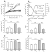
Figure 1
Figure 1
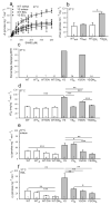
Effect of AICAR on heat induced sudden death in YS mice heat challenged at 37 °C. (a) Oxygen consumption (VO2) during a 15 min exposure of mice at 37 °C, (n = 3–9). (b) VO2 and survival rate (%) of mice exposed to 37 °C as a function of AICAR dose, (n = 3–6). (c) CO2 elimination (VCO2) at the 10th min of heat challenge. (d) Respiratory exchange ratio (RER), calculated as VCO2eliminated/VO2consumed at the 10th min of heat challenge. (e) Serum K+ after 10 min exposure to heat. (f) Rectal temperature, measured immediately after 10 min exposure to heat. In panels c–f AICAR treatment is indicated with subscript A and n numbers are shown. *P < 0.05, **P < 0.01, ***P < 0.001.
Figure 2
Figure 2
AICAR rescue of the YS mice is independent of AMPK activation. (a) Initial phosphorylation rate of SAMS peptide in muscle homogenates. Solid lines represent hyperbolic fits (ν0=(Vmax*[SAMS])/(Km +[SAMS])). Km values are: 145 ± 38 μM (n = 4) for WT soleus, 120 ± 44 μM (n = 3) for YS soleus; 137 ± 37 μM (n = 3) for WT EDL and 149 ± 36 μM (n = 3) for YS EDL. (b) Maximum phosphorylation rate (Vmax) of SAMS peptide in muscle homogenates of soleus (sol) and EDL. (c) Cumulative summary of % of mice of each genotype and treatment that undergo EHR and (d) correspondent VO2 consumption at the 10th min of heat challenge at 37 °C. (e) AMPK activity in soleus and (f) in EDL muscle of mice exposed to 37 °C. AICAR treatment, in panels e–h, is indicated with subscript A, and n numbers are shown in panels d–h. *P < 0.05, **P < 0.01, ***P < 0.001.
Figure 3
Figure 3
Effect of AICAR on RyR1 in the presence of AMP-PCP. (a) Representative plots of 3H-ryanodine binding to WT and YS sarcoplasmic reticulum membranes with increasing AICAR concentrations in the absence of AMP-PCP. (b) Representative plots of 3H-ryanodine binding to WT sarcoplasmic reticulum membranes with increasing concentrations of AMP-PCP in the absence or presence(+A) of 1 mM AICAR. EC50 values: WT 100 ± 5 μM, (n = 3); WT + A 318 ± 51μM, (n = 3), P < 0.001. (c) Representative plots of 3H-ryanodine binding to YS sarcoplasmic reticulum membranes with increasing concentrations of AMP-PCP in the absence or presence(+A) of 1 mM AICAR. EC50 values: YS 153 ± 5 μM; YS+A 330 ± 27 μM, (n = 3), P < 0.01. EC50 values represent the mean of three independent preparations. (d) Representative single channel recordings of WT RyR1 in the presence of 1 mM AMP-PCP, before and after addition of 1 mM AICAR. (e) One subclass of single YS RyR1 channels (others in Supplementary Fig. 4) in the presence of 1 mM AMP-PCP, before and after 1 mM AICAR. (f) RyR1 probability of opening (PO), (g) mean channel open time (τopen) and (h) mean channel closed time (τclosed) for the WT and YS channels. AICAR treatment is indicated with the subscript A, and n numbers are indicated in panels f–h. ***P < 0.001.
Figure 4
Figure 4
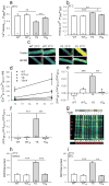
Effect of AICAR on Ca2+, ROS and RNS in single isolated WT and YS FDB fibers. (a) Peak of Ca2+ transient triggered by in vitro application of 4-cmc. (b) Resting cytosolic Ca2+ in indicated fibers. (c) Representative images of single fibers loaded with Fura-2AM. Scale bars represent 20μm. Vertical linear scales represent free [Ca2+] 0–1.7μM (d) Estimation of the changes in resting Ca2+ (nM) with temperature. In vitro AICAR treatment is indicated (+A), (n = 4–6). (e) DAF fluorescence ratio as a measure of RNS production and (f) DCF fluorescence ratio as a measure of ROS production, in FDB fibers. (g) Representative Oxyblot (top) and Coomassie stained gel (bottom) to assess oxidative stress by immunodetection of carbonyl groups (anti-DNP, green) normalized to a non specific band (red) in WT (W) and YS (Y) muscle homogenates. Mice were injected either with saline (−) or AICAR(+) and were heat challenged at 37 °C (+) or not (−) (h) Oxidative stress in soleus and (i) EDL muscle homogenates, as quantified by Oxyblot densitometry. In vitro AICAR application is indicated with subscript A in panels a, b, e, f, h and i, and n numbers are also indicated. *P < 0.05, **P < 0.01, ***P < 0.001.
Figure 5
Figure 5
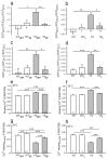
Effects of NOX or NOS inhibition in single isolated FDB fibers at 35 °C. WT and YS fibers were preincubated either with the NOX inhibitor gp91ds-tat peptide (gp) using the corresponding scrambled peptide as control (gps), or the NOS blocker L-NAME (L), blockers are shown as subscripts. DCF fluorescence ratio as a measure of ROS production in fibers incubated with (a) gp91ds-tat peptide or the control peptide and (b) L-NAME. DAF fluorescence ratio as a measure of RNS production, in fibers incubated with (c) gp91ds-tat peptide or the control peptide and (d) L-NAME. Fura-2 ratio as a measure of resting Ca2+ in fibers incubated with (e) gp91ds-tat peptide or the control peptide and (f) L-NAME. Fura-2 ratio as a measure of Ca2+ transient peak triggered by in vitro application of 4-cmc in fibers incubated with (g) gp91ds-tat peptide or the control peptide and (h) L-NAME. The n numbers are indicated in all panels. *P < 0.05, **P < 0.01, ***P < 0.001.
Figure 6
Figure 6
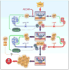
Model for the AICAR prevention of EHR. We are proposing that the heat induced sudden death or EHR in the YS mice arises from a mutation in RyR1 that increases Ca2+ leak at elevated temperatures. The increased cytosolic Ca2+ activates NOX (and to a lesser extent mitochondria) and NOS to produce ROS and RNS, respectively. Both ROS and RNS modify RYR1, and other skeletal muscle proteins to further increase the temperature dependent Ca2+ leak, promoting a feed forward cycle that eventually results in sustained contractures and death. AICAR binds directly to RyR1 to inhibit Ca2+ leak in the presence of cellular concentrations of ATP. Decreased Ca2+ leak prevents ROS and RNS overproduction and stops the feed forward cycle. A role for Ca2+ influx either via CaV1.1, stretch or store operated channels remains to be investigated.

References

    1. Bouchama A, Knochel JP. Heat Stroke. N Engl J Med. 2002;346(25):1978–88. - PubMed
    1. Hopkins PM, Ellis FR, Halsall PJ. Evidence for related myopathies in exertional heat stroke and malignant hyperthermia. The Lancet. 1991;338:1491–92. - PubMed
    1. Jurkat-Rott K, McCarthy T, Lehmann-Horn F. Genetics and Pathogenesis of Malignant Hyperthermia. Muscle Nerve. 2000;23(1):4–17. - PubMed
    1. Wappler F, Fiege M, Steinfath M, Agarwal K, Scholz J, Singh S, Matschke J, Schulte Am Esch J. Evidence for Susceptibility to Malignant Hyperthermia in Patients with Exercise-Induced Rhabdomyolysis. Anesthesiology. 2001;94(1):95–100. - PubMed
    1. Davis M, Brown R, Dickson A, Horton H, James D, Laing N, Marston R, Norgate M, Perlman D, Pollock N, Stonwell K. Malignant Hyperthermia Associated with Exercise-Induced Rhabdomyolysis or Congenital Abnormalities and a Novel RYR1 Mutation in New Zealand and Australian Pedigrees. Br J Anaesth. 2002;88(4):508–15. - PubMed

Publication types

MeSH terms