

doi: 10.1038/nmeth.1841.
Dual-objective STORM reveals three-dimensional filament organization in the actin cytoskeleton
Affiliations
- PMID: 22231642
- PMCID: PMC3304438
- DOI: 10.1038/nmeth.1841
Item in Clipboard
Dual-objective STORM reveals three-dimensional filament organization in the actin cytoskeleton
Nat Methods.
.
Abstract
By combining astigmatism imaging with a dual-objective scheme, we improved the image resolution of stochastic optical reconstruction microscopy (STORM) and obtained <10-nm lateral resolution and <20-nm axial resolution when imaging biological specimens. Using this approach, we resolved individual actin filaments in cells and revealed three-dimensional ultrastructure of the actin cytoskeleton. We observed two vertically separated layers of actin networks with distinct structural organizations in sheet-like cell protrusions.
Conflict of interest statement
The authors declare that they have no competing financial interests.
Figures
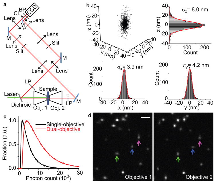
(a) Schematic of the setup. Two microscope objectives are placed opposing each other and focused on the same spot of the sample. Astigmatism is introduced into the images collected by both objectives using a cylindrical lens. M: mirror; Obj.: objective; LP: long-pass filter; CL: cylindrical lens; BP: band-pass filter. (b) Localization precision of Alexa 647 molecules in fixed cells measured with dual-objective STORM. Each molecule gives a cluster of localizations due to repetitive activation of the same molecule. Localizations from 108 clusters (each containing >10 localizations) are aligned by their center of mass to generate the 3D presentation of the localization distribution. Histograms of the distribution in x, y, and z are fit to Gaussian functions, and the resultant standard deviations (σx, σy, and σz) are given in the plots. (c) Distribution of the number of photons detected for individual Alexa 647 molecules through both objectives (red; average: 10,600) and from a single objective (black; average: 5,200). (d) Images of activated Alexa 647 molecules obtained from the two objectives in a single frame. A molecule that appears elongated in x through one objective should appear elongated in y through the opposing objective (green and blue arrows). On the other hand, if two nearby molecules were mistaken as a single molecule, the images obtained through both objectives would appear elongated in the same direction along the line that connects the two molecules (example marked by the magenta arrow). Scale bar: 2 μm.
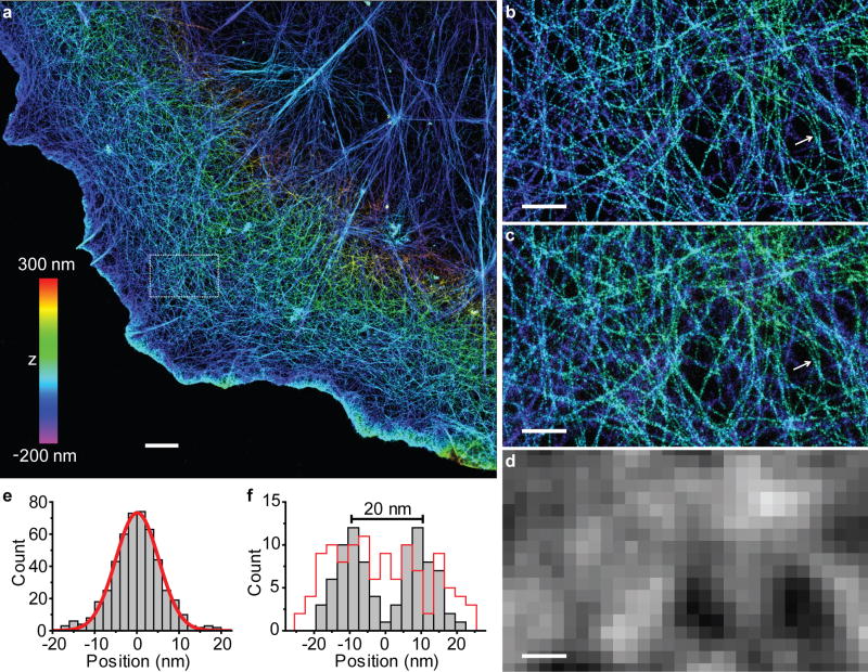
(a) Dual-objective STORM image of actin (labeled with Alexa 647-phalloidin) in a COS-7 cell. The z-positions are color-coded according to the color bar, with violet indicating positions closest to the substratum and red indicating farthest. (b) Close-up of the boxed region in a. (c) STORM image of the same area obtained by using only the information collected by Objective 1 of the dual-objective setup. (d) Conventional fluorescence image of the same area. (e) The cross-sectional profile of eight filaments overlaid by the center of each filament. The red line is a Gaussian fit with FWHM of 12 nm. (f) The cross-sectional profiles for two nearby filaments in (b) and (c) (white arrows). The grey bars correspond to the dual-objective images in (b) and the red line corresponds to the single-objective image in (c). Scale bars: 2 μm for a, 500 nm for b–d.
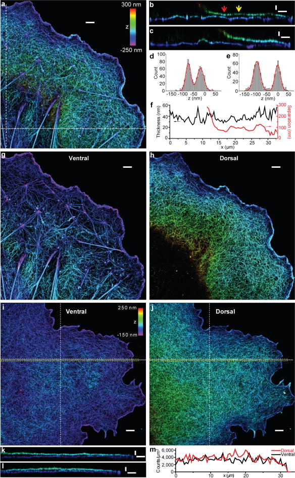
(a) Dual-objective STORM image of actin in a BSC-1 cell. The z-positions are color-coded according to the color bar. (b,c) Vertical cross sections (each 500-nm wide in x or y) of the cell in (a) along the dot and dash lines, respectively. Note when far from the cell edge, the z-position of the dorsal layer increases quickly and falls out of the imaging range. (d,e) The z-profiles for two points along the vertical section, corresponding to the red and yellow arrows in (b), respectively. Each histogram is fit to two Gaussians (red curves), yielding the apparent thickness of the ventral and dorsal layers and the peak separation between the two layers. (f) Quantification of the apparent thickness averaged over the two layers and the dorsal-ventral separation obtained from the xz cross-section profile in (b). (g,h) The ventral and dorsal actin layers of the cell in (a). (i,j) The ventral and dorsal actin layers of a COS-7 cell that was treated with blebbistatin. (k,l) Vertical cross sections (each 500-nm wide in x or y) of the cell along the dot and dash lines, respectively. (m) Actin density of the ventral and dorsal layers along the yellow box in (i,j), measured by the localization density. Scale bars: 2 μm for a, g, h, i, and j; 100 nm for z and 2 μm for x/y for b, c, k and l.
References
Publication types
MeSH terms
Substances
Grants and funding
LinkOut - more resources
Full Text Sources
Other Literature Sources
