

Molecular mechanisms underlying delayed apoptosis in neutrophils from multiple trauma patients with and without sepsis
- PMID: 22231730
- PMCID: PMC3356414
- DOI: 10.2119/molmed.2011.00380
Molecular mechanisms underlying delayed apoptosis in neutrophils from multiple trauma patients with and without sepsis
Abstract
Delayed neutrophil apoptosis and overshooting neutrophil activity contribute to organ dysfunction and subsequent organ failure in sepsis. Here, we investigated apoptotic signaling pathways that are involved in the inhibition of spontaneous apoptosis in neutrophils isolated from major trauma patients with uneventful outcome as well as in those with sepsis development. DNA fragmentation in peripheral blood neutrophils showed an inverse correlation with the organ dysfunction at d 10 after trauma in all patients, supporting the important role of neutrophil apoptosis regulation for patient's outcome. The expression of the antiapoptotic Bcl-2 protein members A1 and Mcl-1 were found to be diminished in the septic patients at d 5 and d 10 after trauma. This decrease was also linked to an impaired intrinsic apoptosis resistance, which has been previously shown to occur in neutrophils during systemic inflammation. In patients with sepsis development, delayed neutrophil apoptosis was found to be associated with a disturbed extrinsic pathway, as demonstrated by reduced caspase-8 activity and Bid truncation. Notably, the expression of Dad1 protein, which is involved in protein N-glycosylation, was significantly increased in septic patients at d 10 after trauma. Taken together, our data demonstrate that neutrophil apoptosis is regulated by both the intrinsic and extrinsic pathway, depending on patient's outcome. These findings might provide a molecular basis for new strategies targeting cell death pathways in apoptosis-resistant neutrophils during systemic inflammation.
Figures
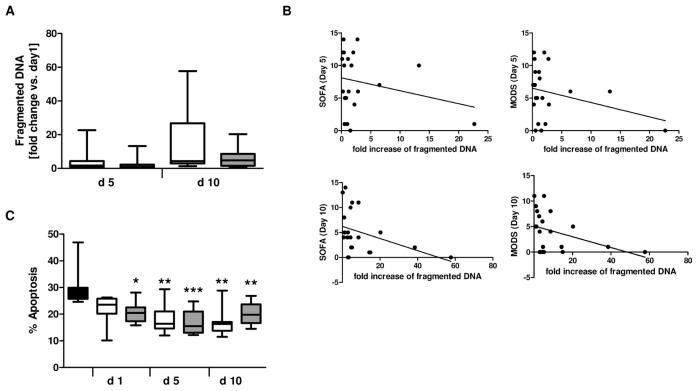
 , sepsis; ■, control. B: SOFA d 5: ρ = −0.23, P = 0.316; SOFA d 10: ρ = −0.417, P = 0.059; MODS d 5: ρ = −0.24, P = 0.29; MODS d 10: ρ = −0.435, P = 0.04.
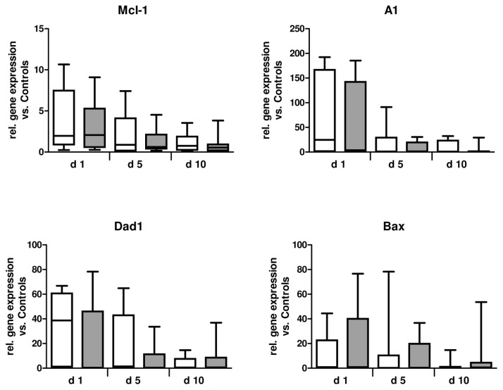 , sepsis.
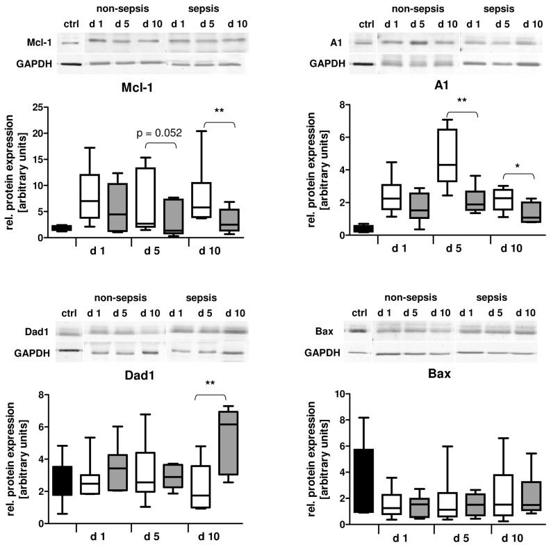 , sepsis; ■, control.
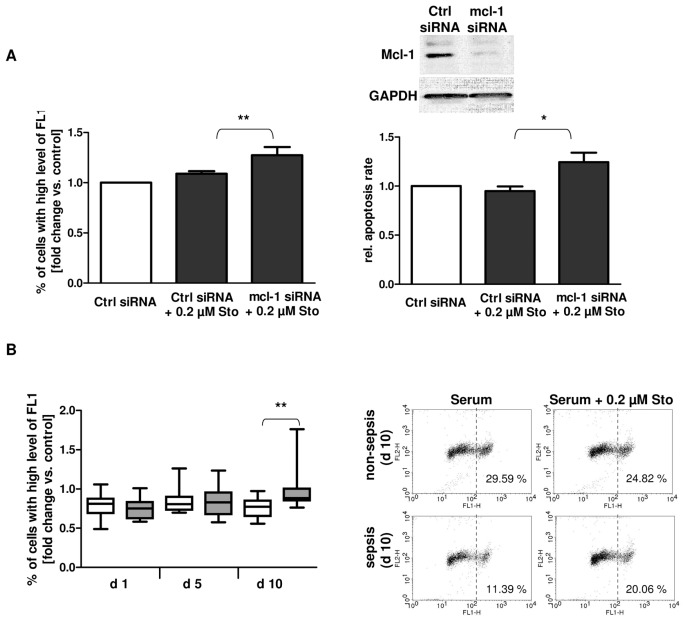 , sepsis.
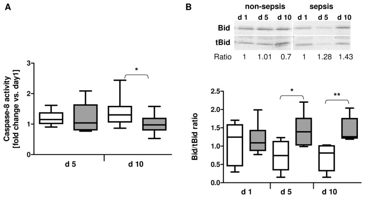 , sepsis.
References
-
- Angus DC, et al. Epidemiology of severe sepsis in the United States: analysis of incidence, outcome, and associated costs of care. Crit Care Med. 2001;29:1303–10. - PubMed
-
- Papathanassoglou ED, Moynihan JA, McDermott MP, Ackerman MH. Expression of Fas (CD95) and Fas ligand on peripheral blood mononuclear cells in critical illness and association with multiorgan dysfunction severity and survival. Crit Care Med. 2001;29:709–18. - PubMed
-
- Power C, Fanning N, Redmond HP. Cellular apoptosis and organ injury in sepsis: a review. Shock. 2002;18:197–211. - PubMed
-
- Vaki I, et al. An early circulating factor in severe sepsis modulates apoptosis of monocytes and lymphocytes. J Leukoc Biol. 2011;89:343–9. - PubMed
-
- Hotchkiss RS, Tinsley KW, Karl IE. Role of apoptotic cell death in sepsis. Scand J Infect Dis. 2003;35:585–92. - PubMed
Publication types
MeSH terms
Substances
LinkOut - more resources
Full Text Sources
Medical
Research Materials
