Review
. 2012 Mar-Apr;4(2):141-62.
doi: 10.1002/wsbm.166. Epub 2012 Jan 9.

Mass spectrometry-based proteomics: qualitative identification to activity-based protein profiling

Affiliations
Review

Mass spectrometry-based proteomics: qualitative identification to activity-based protein profiling

Job D Cardoza et al. Wiley Interdiscip Rev Syst Biol Med. 2012 Mar-Apr.

Abstract

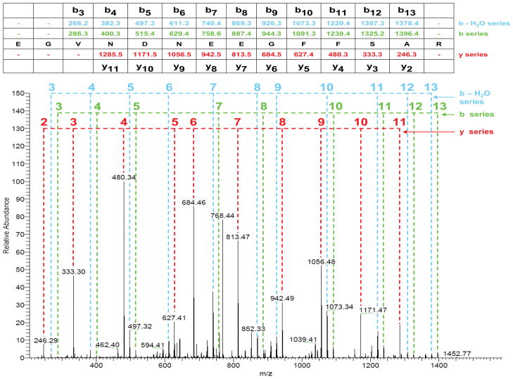
Mass spectrometry has become the method of choice for proteome characterization, including multicomponent protein complexes (typically tens to hundreds of proteins) and total protein expression (up to tens of thousands of proteins), in biological samples. Qualitative sequence assignment based on MS/MS spectra is relatively well-defined, while statistical metrics for relative quantification have not completely stabilized. Nonetheless, proteomics studies have progressed to the point whereby various gene-, pathway-, or network-oriented computational frameworks may be used to place mass spectrometry data into biological context. Despite this progress, the dynamic range of protein expression remains a significant hurdle, and impedes comprehensive proteome analysis. Methods designed to enrich specific protein classes have emerged as an effective means to characterize enzymes or other catalytically active proteins that are otherwise difficult to detect in typical discovery mode proteomics experiments. Collectively, these approaches will facilitate identification of biomarkers and pathways relevant to diagnosis and treatment of human disease.


Figures

Figure 1
Figure 1. LTQ-Orbitrap Mass Spectra for De Novo Sequence Assignment
(A) Example MS spectrum with an m/z window of 300 – 2000 Da. The inset shows the expansion of a 1.5 Da m/z window around the precursor at 785.84 Da. Isotope ions of the precursor are separated by 0.5 Da, indicating a 2+ charge state. (B) MS/MS spectrum for the precursor at m/z = 785.84. The relative intensities of all b- and y-type fragment ions are normalized to the most abundant fragment at m/z = 480.
Figure 2
Figure 2. Peptide Fragmentation Products and Mechanism
(A) Molecular structure of a peptide, with amino acid side chains represented as R1, R2, etc. The fragmentation sites for common ion pairs (a/x, b/y and c/z) are indicated with dashed red lines. The b/y pair is the most commonly observed in CAD mass spectrometry. (B) Mechanism for generation of complimentary b- and y- type ions resulting from fragmentation of a doubly-charged tryptic peptide. Protonation of a backbone amide nitrogen leads to lysis of the amide bond and formation of a cyclized oxazolone ion structure, along with the corresponding y-type ion. The oxazolone ring on the b ion can undergo electron rearrangement, leading to loss of a carbonyl group and formation of an a-type fragment ion. *The illustrations in this figure were generated using ACD/Chemsketch Freeware, version 12.01, Advanced Chemistry Development, Inc., Toronto, ON, Canada, www.acdlabs.com, 2010.
Figure 3
Figure 3. Fragment ion Assignment in an MS/MS Spectrum
The MS/MS spectrum for the 785.84 Da, 2+ precursor in Fig. 1B is shown with the b- and y- type ions assigned. In addition, the b – H2O ion series is also labeled. The fragment ion masses for each series are shown in the data table above the spectrum. The b and b – H2O ion series always contain the N-terminus and hence progress from the N- to C-terminus. In contrast the y-type ions contain the C-terminus and progress in the opposite direction. For clarity the b6 and b9 ion labels have been omitted from the spectrum due to their proximity to the y5 and y8 ions, respectively, though they are present and their masses are shown in the data table.
Figure 4
Figure 4. Enrichment of Biological Annotations
An example enrichment analysis displays signaling pathways from the KEGG database whose members are significantly enriched within an experimental dataset. Top: Enrichment analysis involves quantifying the overlap between experimentally determined protein lists (green circles) and protein sets for a particular biological theme extracted from databases such as KEGG (blue circles). The significance of enrichment, computed as a p-value based on a Fisher’s Exact Test, accounts for the degree of overlap between an empirically-determined protein list and the protein set corresponding to a particular KEGG pathway, in addition to the size of the input protein list, KEGG pathway protein set, and the total proteome. Bottom: In this example, KEGG signaling pathways that score as “enriched” (p-value < 0.05) are displayed as a bar graph.
Figure 5
Figure 5. Networks
Networks are often used to visualize data where nodes represent experimentally determined proteins and edges represent biochemical relationships extracted from publicly accessible databases. (A) The most common approach is to connect proteins using undirected protein-protein interaction edges (in gray) and to (B) color the nodes based on quantitative data (e.g., “upregulated or downregulated”). In this example, red and blue nodes indicate increased and reduced levels of phosphorylation, respectively. (C) Directed networks are often more appropriate, especially when representing enzymatic and other transient relationships. Here, edges terminated with purple arrows and brown circles represent kinase- and phosphatase- substrate relationships, respectively. Networks lend themselves to application of graph theoretical methods such as the (D) Steiner Tree algorithm, which introduces undetected nodes (yellow) in order to connect all experimental nodes using a minimal number of edges. Integration of multiple edge types and graph theory algorithms can provide a more comprehensive description of signaling pathways.
Figure 6
Figure 6. Activity-based Protein Profiling – ABPP
(A) Essential components of an activity-based probe include a reactive group (RG) that targets a particular enzyme class as well as a reporter group (Rp) that allows for isolation and/or detection of labeled molecules. (B) The copper-catalyzed click chemistry reaction between an azide and an alkyne can be used to couple reactive probes to reporter functionalities in lysates after labeling in vivo.
Figure 7
Figure 7. Mass Spectrometry-based Strategies to Identify Proteins Captured by Activity Probes
Affinity tags (e.g., biotin) are typically required to facilitate enrichment of proteins targeted by activity-based probes. Depending on the analytical strategy, MS interrogation can be used to (A) sequence proteolytic peptides from labeled proteins, (B) detect probe modified peptides, or (C) detect both in separate LC-MS/MS analyses.

References

    1. Weston AD, Hood L. Systems biology, proteomics, and the future of health care: toward predictive, preventative, and personalized medicine. J Proteome Res. 2004;3(2):179–96. - PubMed
    1. Hu Q, et al. The Orbitrap: a new mass spectrometer. J Mass Spectrom. 2005;40(4):430–443. - PubMed
    1. Loboda AV, et al. A tandem quadrupole/time-of-flight mass spectrometer with a matrix-assisted laser desorption/ionization source: design and performance. Rapid Commun Mass Spectrom. 2000;14(12):1047–57. - PubMed
    1. Wenzel RJ, et al. Analysis of megadalton ions using cryodetection MALDI time-of-flight mass spectrometry. Anal Chem. 2005;77(14):4329–37. - PubMed
    1. Zhou M, Robinson CV. When proteomics meets structural biology. Trends Biochem Sci. 2010;35(9):522–9. - PubMed

Publication types