. 2012 Mar;80(3):929-42.
doi: 10.1128/IAI.05769-11. Epub 2012 Jan 9.

Hemin binding protein C is found in outer membrane vesicles and protects Bartonella henselae against toxic concentrations of hemin

Affiliations

Hemin binding protein C is found in outer membrane vesicles and protects Bartonella henselae against toxic concentrations of hemin

Julie A Roden et al. Infect Immun. 2012 Mar.

Abstract

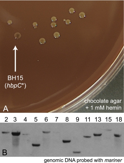
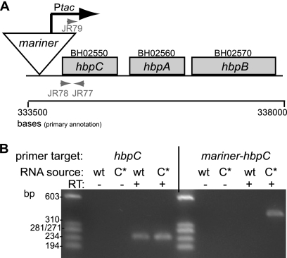
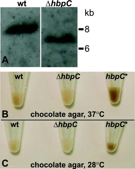
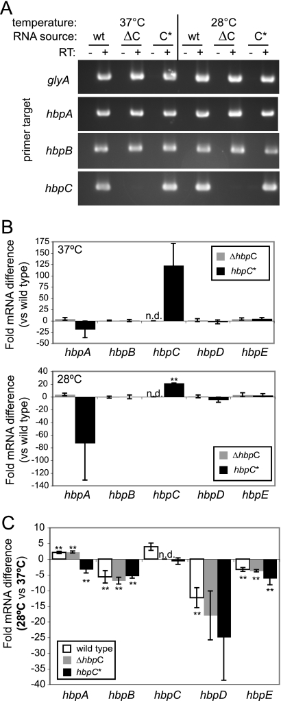
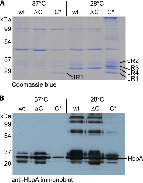
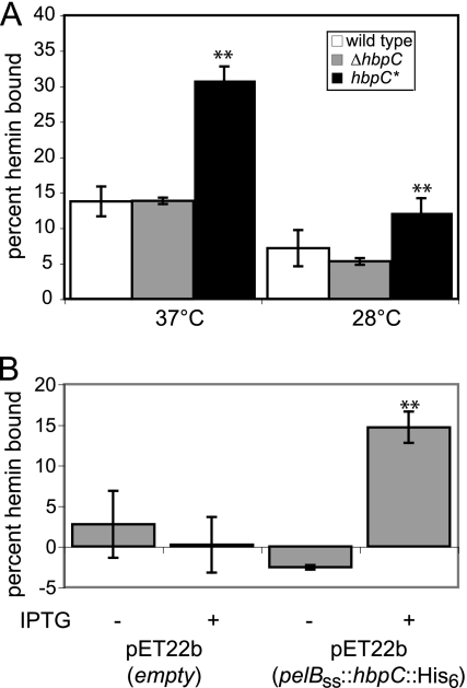
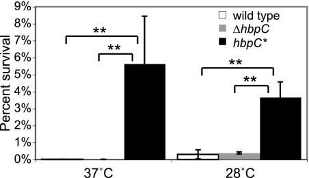
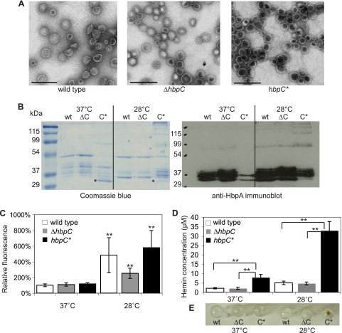
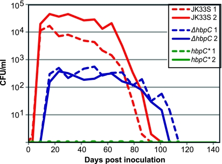
Bartonella species are gram-negative, emerging bacterial pathogens found in two distinct environments. In the gut of the obligately hematophagous arthropod vector, bartonellae are exposed to concentrations of heme that are toxic to other bacteria. In the bloodstream of the mammalian host, access to heme and iron is severely restricted. Bartonellae have unusually high requirements for heme, which is their only utilizable source of iron. Although heme is essential for Bartonella survival, little is known about genes involved in heme acquisition and detoxification. We developed a strategy for high-efficiency transposon mutagenesis to screen for genes in B. henselae heme binding and uptake pathways. We identified a B. henselae transposon mutant that constitutively expresses the hemin binding protein C (hbpC) gene. In the wild-type strain, transcription of B. henselae hbpC was upregulated at arthropod temperature (28°C), compared to mammalian temperature (37°C). In the mutant strain, temperature-dependent regulation was absent. We demonstrated that HbpC binds hemin and localizes to the B. henselae outer membrane and outer membrane vesicles. Overexpression of hbpC in B. henselae increased resistance to heme toxicity, implicating HbpC in protection of B. henselae from the toxic levels of heme present in the gut of the arthropod vector. Experimental inoculation of cats with B. henselae strains demonstrated that both constitutive expression and deletion of hbpC affect the ability of B. henselae to infect the cat host. Modulation of hbpC expression appears to be a strategy employed by B. henselae to survive in the arthropod vector and the mammalian host.


Figures

Fig 1
Fig 1
mariner transposon mutagenesis of B. henselae JK33S yields strains with darker pigmentation than that of the wild-type parent. (A) Dark colonies, such as BH15, are easily observed with a visual screen of mutagenized Bartonella colonies grown on chocolate agar supplemented with 1 mM hemin. (B) The mariner transposon inserts once per strain, as demonstrated by Southern blotting of genomic DNA from transposon-mutagenized bacteria, using the pBT20 mariner sequence as a probe.
Fig 2
Fig 2
Insertion of the mariner transposon construct pBT20 upstream of a locus containing three hemin binding protein (hbp) genes in mutant strain BH15 (hbpC*) yields constitutive expression of hbpC. (A) Sequencing of BH15 shows that the mariner insertion site in this mutant strain is located directly upstream of hbpC. Designations above each hbp represent primary open reading frame (ORF) annotations. (B) The constitutive expression of hbpC in B. henselae JK33S hbpC* is the result of transcription from the mariner Ptac promoter. Internal hbpC primers (JR78 and JR77) amplify a 242-bp product from both JK33S (wild type [wt]) and JK33S hbpC* (C*) cDNA, whereas the Ptac-hbpC primer pair (JR79 and JR77) produces only a 399-bp product from JK33S hbpC* cDNA. RT indicates reverse transcriptase.
Fig 3
Fig 3
Generation of a B. henselae JK33S hbpC null mutant and comparison of pigmentation of the three isogenic strains JK33S wild type, JK33S ΔhbpC, and JK33S hbpC*. (A) A Southern blot of genomic DNA extracted from individual B. henselae JK33S (wt) and B. henselae ΔhbpC colonies was probed with the fused fragments from the genomic regions 5′ and 3′ to hbpC, confirming construction of a ΔhbpC mutant. (B and C) To examine pigmentation differences due to the presence or absence of hbpC, all 3 strains of B. henselae were grown on chocolate agar at 37°C or 28°C. The ΔhbpC mutant has pigmentation similar to that of the JK33S wild-type strain, whereas the hbpC* strain demonstrates a hyperpigmentation phenotype at both temperatures.
Fig 4
Fig 4
Constitutive transcription or deletion of hbpC alters transcription of other hbp genes. (A) hbpA and hbpB are transcribed in the B. henselae JK33S ΔhbpC mutant grown at 28°C and 37°C. No hbpC mRNA is detected in B. henselae JK33S ΔhbpC. The reference gene glyA was amplified as a control for RT-PCR. (B) RT-qPCR was used to determine the fold change in mRNA level for each hbp gene in the ΔhbpC and hbpC* strains compared to the level of the same hbp gene in the B. henselae JK33S wild-type strain. mRNA levels of hbpC are increased and mRNA levels of hbpA are decreased in B. henselae JK33S hbpC* grown at both temperatures, compared to the levels for wild-type JK33S. (C) Transcript levels of each hbp gene at 28°C were compared with mRNA levels of the same hbp gene from the same strain grown at 37°C. hbpA and hbpC mRNA levels are increased in the wild-type strain JK33S and hbpB, hbpD, and hbpE transcript levels are decreased in all three strains grown at 28°C, compared with results at 37°C. Error bars show standard errors of the means for three independent experiments and RNA preparations. For each independent experiment, each variable was performed in triplicate. RT indicates reverse transcriptase, n.d. indicates that values for these samples were not determined, and ** indicates a significant difference between mRNA fold change levels in the two strains compared (two-tailed t test, P < 0.05).
Fig 5
Fig 5
Subcellular fractionation of B. henselae JK33S, JK33S ΔhbpC, and JK33S hbpC* protein lysates shows that constitutive expression of hbpC at 28°C and 37°C results in increased HbpC in the outer membrane. (A) After Sarkosyl-based subcellular fractionation of JK33S (wt), JK33S ΔhbpC (ΔC), and JK33S hbpC* (C*) strains, equal amounts of OMP were loaded, separated by SDS-PAGE, and visualized by Coomassie blue staining. The constitutively expressed OMP JR1, present in the B. henselae hbpC* strain at 37°C and 28°C, was identified as HbpC by mass spectrometry (Table 4). The proteins JR2, JR3, and JR4, which are evident in the OMP fraction of all three strains at 28°C, were identified as HbpA (JR2), HbpA and ComL (JR3), and HbpA (JR4) (Table 4). (B) Immunoblotting of the OMP subcellular fractions using anti-HbpA antibodies demonstrated that HbpA expression is substantially upregulated in B. henselae wt and ΔhbpC strains at 28°C compared with that at 37°C. In contrast, HbpA expression is downregulated at both 28°C and 37°C when hbpC is constitutively expressed.
Fig 6
Fig 6
Overexpression of HbpC in B. henselae JK33S hbpC* or E. coli BL21(DE3) significantly increases hemin binding capacity compared with that for control strains. (A) B. henselae cells were incubated with 30 μg/ml (∼0.05 mM) hemin for 1 h. The JK33S hbpC* strain, which constitutively expresses HbpC, shows a significant increase in the amount of hemin bound, compared to that for the JK33S wild-type and JK33S ΔhbpC strains, at 28°C and 37°C. (B) E. coli BL21(DE3) strains carrying pET22b or pET22b(pelBSS::hbpC::His6) were induced at 37°C with 1 mM IPTG to express PelBSS::HbpC::His6 and assayed for hemin binding. Heterologous expression of HbpC in E. coli confers the ability to bind significantly more hemin than the induced, empty-vector control. For both B. henselae and E. coli experiments, the amount of hemin bound was calculated and compared to that for a control reaction mixture without bacteria. Data shown are from one representative experiment with assays performed in triplicate. Error bars represent 95% confidence intervals. ** indicates a significant difference in results between B. henselae JK33S hbpC* and the other two B. henselae strains or between induced and uninduced E. coli strains (two-tailed t test, P < 0.05).
Fig 7
Fig 7
Constitutive expression of hbpC protects B. henselae against hemin toxicity. B. henselae JK33S, JK33S ΔhbpC, and JK33S hbpC* were incubated with 5 mM hemin or a pH-identical control solution in M199S medium for 24 h, and surviving bacterial cells were enumerated. Percent survival after hemin exposure was calculated by comparing the number of viable colonies after incubation with hemin to the number of viable colonies after incubation in the control medium. A significantly higher percentage of the B. henselae bacteria that constitutively express hbpC survived when incubated at either 37°C or 28°C. Assays were performed in triplicate. Data shown are from one representative experiment. Error bars represent 95% confidence intervals. ** indicates a significant difference (two-tailed t test, P < 0.05).
Fig 8
Fig 8
B. henselae produces outer membrane vesicles (OMV) that contain HbpA and HbpC, and constitutive expression of HbpC increases the amount of hemin associated with OMV. (A) OMV isolated from B. henselae JK33S, JK33S ΔhbpC, and JK33S hbpC* grown on chocolate agar were negatively stained with 2% uranyl acetate and visualized by TEM, revealing vesicles between 20 and 100 nm in size. Bar, 500 nm. (B) Proteins present in the OMV were separated by SDS-PAGE and visualized by Coomassie blue staining (left). Equal amounts of total protein were loaded for each strain. HbpC was identified in the OMV fractions from JK33S hbpC* grown at 28°C and 37°C (asterisks); mass spectrometry identified these bands at ∼30 kDa as a mixture of HbpC and HbpA (Table 4, JR5). HbpA was detected in the OMV by immunoblotting each strain grown at both temperatures with anti-HbpA antibodies (right). (C) OMV fractions were incubated with FM 4-64 membrane dye, and relative fluorescence was calculated by assigning the relative fluorescence of B. henselae JK33S grown at 37°C at 100%. Fluorescence values for each strain were compared between OMV fractions from cells grown at 37°C and 28°C. Data shown are from one representative experiment, with assays performed in triplicate. A significantly greater number of OMV is produced by each strain grown at 28°C than by the same strain grown at 37°C. (D) The concentration of hemin associated with OMV was calculated by measuring the absorbance of OMV fractions at 400 nm. Absorbance was measured in triplicate for each OMV sample, and data shown are from one representative experiment. The amount of hemin associated with the JK33S hbpC* OMV is significantly greater than the hemin associated with the JK33S wild type or JK33S ΔhbpC OMV. (E) Images of the OMV pellets generated from bacterial suspensions of equivalent OD demonstrate increased pigmentation of the OMV from strain JK33S hbpC* grown at 28°C, due to the increased association of hemin with this strain. Error bars represent 95% confidence intervals. ** indicates a significant difference (two-tailed t test, P < 0.05).
Fig 9
Fig 9
B. henselae JK33S, JK33S ΔhbpC, and JK33S hbpC* demonstrate differing levels of virulence in vivo in the feline natural host. Two cats each were inoculated intradermally with 2 × 10⁸ CFU of one of the 3 bacterial strains, and blood was cultured for B. henselae for 140 days. No colonies were isolated from either cat infected with JK33S hbpC* throughout the course of infection, and the maximal titers from the two cats inoculated with JK33S ΔhbpC were 100-fold lower than those from the two cats inoculated with the JK33S wild type.

Similar articles

Cited by

References

    1. Alsmark CM, et al. 2004. The louse-borne human pathogen Bartonella quintana is a genomic derivative of the zoonotic agent Bartonella henselae. Proc. Natl. Acad. Sci. U. S. A. 101:9716–9721 - PMC - PubMed
    1. Battisti JM, Sappington KN, Smitherman LS, Parrow NL, Minnick MF. 2006. Environmental signals generate a differential and coordinated expression of the heme receptor gene family of Bartonella quintana. Infect. Immun. 74:3251–3261 - PMC - PubMed
    1. Battisti JM, et al. 2007. Transcriptional regulation of the heme binding protein gene family of Bartonella quintana is accomplished by a novel promoter element and iron response regulator. Infect. Immun. 75:4373–4385 - PMC - PubMed
    1. Bibb LA, Kunkle CA, Schmitt MP. 2007. The ChrA-ChrS and HrrA-HrrS signal transduction systems are required for activation of the hmuO promoter and repression of the hemA promoter in Corynebacterium diphtheriae. Infect. Immun. 75:2421–2431 - PMC - PubMed
    1. Bibb LA, Schmitt MP. 2010. The ABC transporter HrtAB confers resistance to hemin toxicity and is regulated in a hemin-dependent manner by the ChrAS two-component system in Corynebacterium diphtheriae. J. Bacteriol. 192:4606–4617 - PMC - PubMed

Publication types

LinkOut - more resources