. 2012 Feb;122(2):759-76.
doi: 10.1172/JCI57313. Epub 2012 Jan 9.

The CXCR4/CXCR7/SDF-1 pathway contributes to the pathogenesis of Shiga toxin-associated hemolytic uremic syndrome in humans and mice

Affiliations

The CXCR4/CXCR7/SDF-1 pathway contributes to the pathogenesis of Shiga toxin-associated hemolytic uremic syndrome in humans and mice

Tania N Petruzziello-Pellegrini et al. J Clin Invest. 2012 Feb.

Abstract

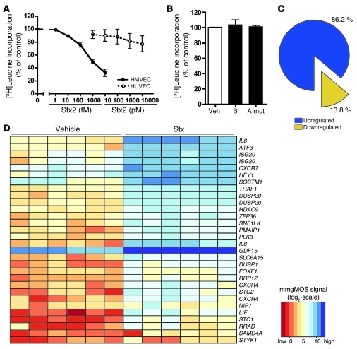
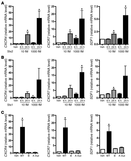
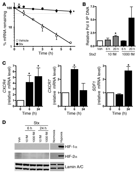
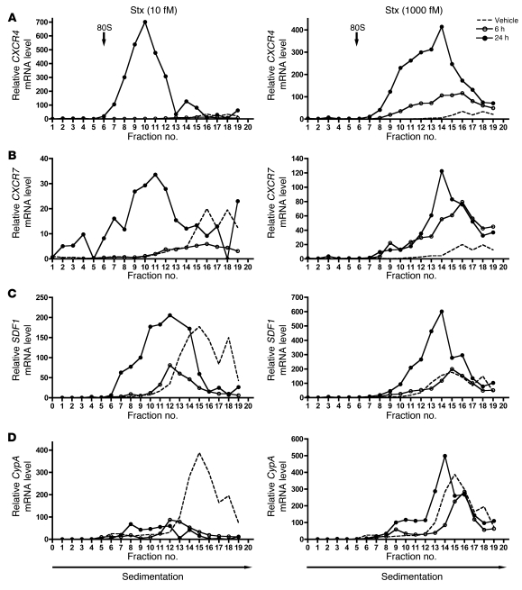
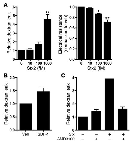
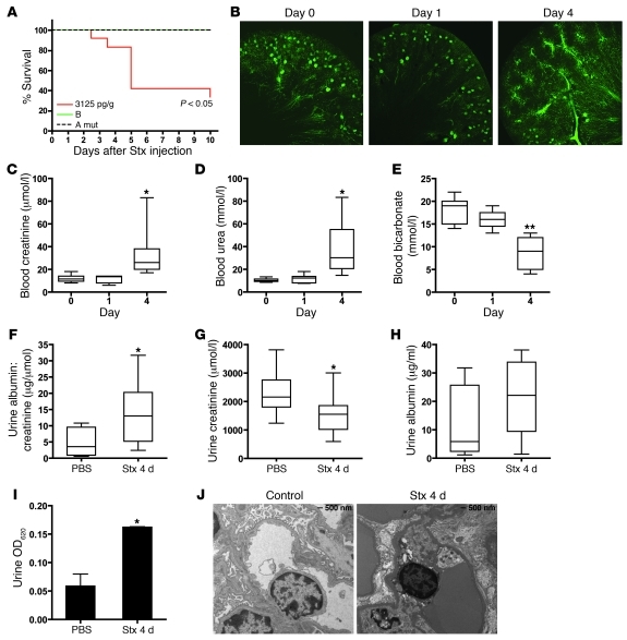
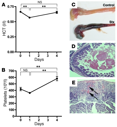
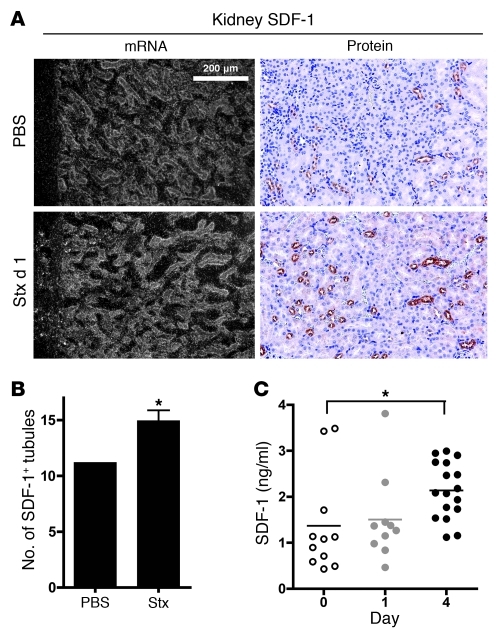
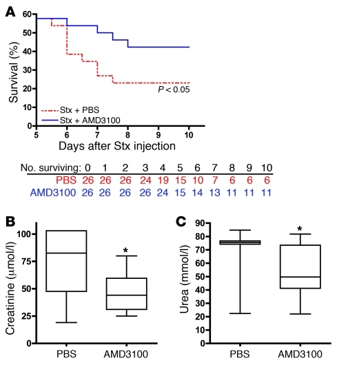
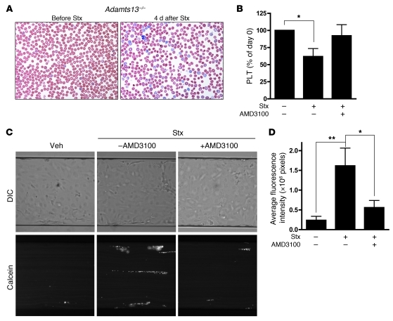
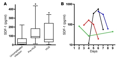
Hemolytic uremic syndrome (HUS) is a potentially life-threatening condition. It often occurs after gastrointestinal infection with E. coli O157:H7, which produces Shiga toxins (Stx) that cause hemolytic anemia, thrombocytopenia, and renal injury. Stx-mediated changes in endothelial phenotype have been linked to the pathogenesis of HUS. Here we report our studies investigating Stx-induced changes in gene expression and their contribution to the pathogenesis of HUS. Stx function by inactivating host ribosomes but can also alter gene expression at concentrations that minimally affect global protein synthesis. Gene expression profiling of human microvascular endothelium treated with Stx implicated a role for activation of CXCR4 and CXCR7 by their shared cognate chemokine ligand (stromal cell-derived factor-1 [SDF-1]) in Stx-mediated pathophysiology. The changes in gene expression required a catalytically active Stx A subunit and were mediated by enhanced transcription and mRNA stability. Stx also enhanced the association of CXCR4, CXCR7, and SDF1 mRNAs with ribosomes. In a mouse model of Stx-mediated pathology, we noted changes in plasma and tissue content of CXCR4, CXCR7, and SDF-1 after Stx exposure. Furthermore, inhibition of the CXCR4/SDF-1 interaction decreased endothelial activation and organ injury and improved animal survival. Finally, in children infected with E. coli O157:H7, plasma SDF-1 levels were elevated in individuals who progressed to HUS. Collectively, these data implicate the CXCR4/CXCR7/SDF-1 pathway in Stx-mediated pathogenesis and suggest novel therapeutic strategies for prevention and/or treatment of complications associated with E. coli O157:H7 infection.


Figures

Figure 1
Figure 1. Stx have potent effects on endothelial cell metabolism and phenotype.
HMVECs or HUVECs were treated with vehicle (Veh) or the indicated concentrations of (A) Stx2, (B) Stx1 B subunit alone (B) (1,000 fM), or holotoxin with a double mutation in the A subunit (A mut) (1,000 fM) for 24 hours. One hour before harvest, 1 μCi [3H]leucine was added to assess de novo protein synthesis. Radioactivity incorporated into TCA-precipitable material was quantitated, and data were normalized to vehicle-treated cells. The mean ± SEM of 14 experiments (HMVECs with Stx2), 5 experiments (HUVECs), or 3 experiments (mutants), triplicate determinations, is shown. (C) HMVECs (n = 6 replicates) were treated with Stx2 (10 fM, 24 hours) or vehicle and subjected to Affymetrix GeneChip gene expression profiling. By combining probe sets identified as differentially expressed by all preprocessing algorithms and removing redundant probe sets, i.e., probe sets mapping to the same gene, it was determined that 86.2% of differentially expressed genes (318 out of 369 unique genes) were upregulated by treatment with Stx. (D) The top 30 differentially expressed probe sets (based on magnitude of differential expression) were summarized in a heat map with hierarchical clustering. Processed signal intensities (computed using the mmgMOS preprocessing algorithm) are depicted on a log2 scale, with rows representing differentially expressed probe sets and columns representing Stx- or vehicle-treated samples. Note that several probe sets correspond to the same genes.
Figure 2
Figure 2. Stx activates the CXCR4/CXCR7/SDF-1 pathway in microvascular endothelium by a mechanism requiring enzymatic activity of the Stx A subunit.
HMVECs were treated with the indicated concentrations of (A) Stx2 or (B) Stx1 for 6 or 24 hours, and mRNA levels were quantitated by qRT-PCR. (C) HMVECs were treated with 1,000 fM wild-type Stx2 (WT), Stx1 B subunit alone, or Stx2 holotoxin consisting of a mutant A subunit and wild-type B subunit (A mut). mRNA levels were measured by qRT-PCR. In all cases, data are normalized for GAPDH and 18S and are shown relative to vehicle-treated cells. Similar trends were observed whether or not data were normalized to housekeeping genes. Basal levels of CXCR4, CXCR7, and SDF1 were 1.53 ± 0.43 × 10⁴, 7.40 ± 2.20 × 10⁴, and 2.21 ± 0.69 × 10⁴ copies/μg of input RNA, respectively. The mean ± SEM of at least 4 independent experiments is shown. *P < 0.05 vs. vehicle-treated cells.
Figure 3
Figure 3. Stx enhances CXCR4 expression by both transcriptional and posttranscriptional mechanisms.
(A) HMVECs treated with vehicle or 1,000 fM Stx2 for 20 hours were subjected to transcription arrest with 10 μg/ml actinomycin D, and RNA was harvested at various times thereafter. Remaining CXCR4 mRNA was determined using qRT-PCR and normalized to 18S. The mean ± SEM of 3 independent experiments is shown. Half-lives were calculated using exponential regression to be 1.18 ± 0.09 hours in vehicle-treated cells and 19.2 ± 7.8 hours in Stx-treated cells. (B) HMVECs were exposed to 1,000 fM Stx2 for 20 hours, followed by ChIP using anti-RNA polymerase II (Pol II) antibodies. qRT-PCR for CXCR4 was then used to determine the amount of immunoprecipitated DNA (IP DNA). The mean ± SEM of at least 4 independent experiments is shown. (C) HMVECs were maintained under normoxic (21% O2) or hypoxic (<1% O2) conditions for the indicated times. qRT-PCR was used to determine mRNA levels. Data are normalized for 18S levels and are shown relative to normoxic cells. The mean ± SEM of 3 independent experiments is shown. Similar trends were observed whether or not data are normalized to housekeeping genes. (D) Western blot analyses were performed for HIF-1α and HIF-2α in HMVECs treated with 10 fM or 1,000 fM Stx for 6 or 24 hours. Lamin A/C was used as a loading control. Negative control and positive control were normoxic and hypoxic HMVECs. *P < 0.05 vs. vehicle or normoxia.
Figure 4
Figure 4. Stx-treated cells exhibit increased CXCR4, CXCR7, and SDF1 transcripts associated with cellular polyribosomes.
HMVECs were treated with vehicle or the indicated concentrations of Stx for 6 or 24 hours. Cellular ribosomes were fractionated based on density into 20 fractions, and RNA was purified from each fraction. mRNA levels were quantitated by qRT-PCR, normalized for luciferase mRNA, and shown relative to vehicle fraction no. 1. 80S refers to 80S ribosome. (A) CXCR4, (B) CXCR7, (C) SDF1, and (D) cyclophilin A. A representative experiment is shown.
Figure 5
Figure 5. Functional effects of Stx and the CXCR4/CXCR7/SDF-1 axis on endothelial permeability.
(A) Confluent endothelial monolayers were grown on transwell filters and treated with the indicated concentrations of Stx. Stx treatment causes increased dextran leak and decreased electrical resistance across the monolayer. The mean ± SEM of at least 3 independent experiments is shown. (B) Treatment of HMVECs with SDF-1 (50 ng/ml, 5 hours) induces dextran leak. The mean ± SEM of at least 3 independent experiments is shown. (C) Stx-induced HMVEC permeability is decreased by treatment of AMD3100/plerixafor. A representative experiment of 3 is shown (mean ± SD). *P < 0.05, **P < 0.001 vs. vehicle-treated cells.
Figure 6
Figure 6. Stx challenge causes impaired renal function and mortality in vivo.
(A) CAST/Ei mice were injected i.v. with wild-type Stx2 (3,125 pg/g), Stx2 A subunit mutant (10 times LD50), or Stx1 B subunit (50 times LD50), and survival was monitored for 10 days. (B) Mice were injected with Stx2 (2,400 pg/g) and, at the indicated times, animals were perfused with fluorescent microbeads. A representative confocal image of a 100-μm section is shown (original magnification, ×5). Plasma collected from Stx-treated animals shows elevated (C) creatinine and (D) urea and reduced (E) bicarbonate levels by 4 days after Stx injection. Mice injected with Stx had (F) significantly higher urine albumin/creatinine ratios by 4 days after Stx injection, (G) lower urine creatinine, and (H) slightly elevated urine albumin. Horizontal bars indicate the median values, boxes indicate 25th to 75th percentiles, and whiskers indicate the minimum and maximum values. (I) Mice injected with Stx or PBS were injected i.v. with 0.1% Evans Blue dye. Urine samples were collected 1 hour later, and the amount of dye leak was measured spectrophotometrically. (J) Transmission electron micrographs of glomeruli from control and Stx-treated animals. *P < 0.05, **P < 0.001 vs. day 0 or PBS control.
Figure 7
Figure 7. Effects of Stx on animal hematology and gastrointestinal biology.
(A) CAST/Ei mice were injected i.v. with Stx2 (2,400 pg/g) on day 0. Blood samples were taken via saphenous vein at the indicated times. (A) Hematocrit (HCT) and (B) platelet levels are shown (n = 13 per time point). (CE) Approximately 60% of mice develop gastrointestinal hemorrhage. (C) Gross images of stomach and small intestine from PBS- and Stx-treated mice. (D) H&E staining shows intraluminal hemorrhage (original magnification, ×50) and (E) intramucosal hemorrhage (arrows; original magnification, ×100). **P < 0.001.
Figure 8
Figure 8. Stx challenge causes dysregulation of SDF-1 in vivo.
(A) SDF-1 in situ hybridization and immunohistochemistry demonstrate increased SDF-1 production in the renal cortical tubules (original magnification, ×160). (B) The number of tubules staining positive for SDF-1 by immunohistochemistry were quantified in 6 nonoverlapping cortical fields (magnification, ×100). (C) Animals were injected with Stx2 (2,400 pg/g) on day 0, and SDF-1α levels were measured in blood plasma at the indicated times. Horizontal bars indicate the mean, and each symbol represents an individual animal. *P < 0.01.
Figure 9
Figure 9. Blockade of CXCR4/SDF-1 interaction by AMD3100 improves renal function and promotes animal survival.
(A) CAST/Ei mice were injected with Stx2 (2,400 pg/g) on day 0, followed by daily administration of AMD3100 (10 μg/g) or an equal volume of PBS beginning 1 day after Stx. Survival was monitored for 10 days (n = 26 per treatment group). (B and C) CAST/Ei mice were injected with Stx2 (625 pg/g) on day 0, followed by daily injections of AMD3100 (10 μg/g) or an equivalent volume of PBS vehicle. Plasma was collected 4 days after Stx injection and analyzed for (B) creatinine and (C) urea (n = 10–26 animals per group). Horizontal bars indicate the median values, boxes indicate 25th to 75th percentiles, and whiskers indicate the minimum and maximum values. *P < 0.05.
Figure 10
Figure 10. Blockade of CXCR4/SDF-1 interaction by AMD3100 restores platelet levels in vivo and inhibits platelet adhesion to endothelial cells under flow conditions in vitro.
(A) Blood smears from Adamts13–/– mice injected with Stx2 (625 pg/g) show evidence of red blood cell fragments 4 days after Stx exposure, with high numbers of reticulocytes (original magnification, ×150). (B) Adamts13–/– mice were injected with Stx2 (625 pg/g) on day 0, followed by daily injections of AMD3100 (10 μg/g) or PBS. Blood was analyzed for platelet levels prior to Stx injection and again at day 4 after toxin injection in both groups (n = 9–21 animals per group). *P < 0.05 vs. control animals that received neither Stx nor AMD3100. (C) HMVECs were grown to confluence in microfluidic channels and were either left untreated or were treated with 10 fM Stx2 (24 hours), after which calcein-labeled platelets were perfused at constant shear rates of 2.5 dyn/cm2 over the endothelial monolayer. Preincubation of HMVECs with AMD3100 (10 μM) significantly inhibited platelet adhesion. Images are representative of 5 independent experiments (original magnification, ×15). (D) Quantification of platelet adhesion to endothelial cells. Data are expressed as mean ± SEM from 5 independent experiments. *P < 0.05; **P < 0.01.
Figure 11
Figure 11. Circulating SDF-1 levels in HUS patients.
SDF-1 levels were measured in plasma samples from individuals infected with E. coli O157:H7. (A) Infected samples obtained from patients with uncomplicated O157:H7 infection within 4 days after the onset of diarrhea (n = 36). Horizontal bars indicate the median values, boxes indicate 25th to 75th percentiles, and whiskers indicate the minimum and maximum values. Pre-HUS, samples collected prior to HUS diagnosis in those individuals who later progressed to HUS (n = 7); HUS, samples collected on the day of HUS diagnosis (n = 22). *P < 0.05. (B) SDF-1 levels over the course of E. coli O157:H7–mediated illness. Each line represents 1 patient, and the last data point for each patient was obtained on the day of HUS diagnosis.

Similar articles

Cited by

References

    1. Karmali MA. Infection by verocytotoxin-producing Escherichia coli. Clin Microbiol Rev. 1989;2(1):15–38. - PMC - PubMed
    1. Bielaszewska M, et al. Characterisation of the Escherichia coli strain associated with an outbreak of haemolytic uraemic syndrome in Germany, 2011: a microbiological study. Lancet Infect Dis. 2011;11(9):671–676. - PubMed
    1. Frank C, et al. Epidemic profile of Shiga-toxin-producing Escherichia coli O104:H4 outbreak in Germany. N Engl J Med. 2011;365(19):1771–1780. doi: 10.1056/NEJMoa1106483. - DOI - PubMed
    1. Rohde H, et al. Open-source genomic analysis of Shiga-toxin-producing E. coli O104:H4. N Engl J Med. 2011;365(8):718–724. doi: 10.1056/NEJMoa1107643. - DOI - PubMed
    1. Rasko DA, et al. Origins of the E. coli strain causing an outbreak of hemolytic-uremic syndrome in Germany. N Engl J Med. 2011;365(8):709–717. doi: 10.1056/NEJMoa1106920. - DOI - PMC - PubMed

Publication types

MeSH terms