Review
. 2012 Jan;25(1):193-213.
doi: 10.1128/CMR.00013-11.

Polymicrobial interactions: impact on pathogenesis and human disease

Affiliations
Review

Polymicrobial interactions: impact on pathogenesis and human disease

Brian M Peters et al. Clin Microbiol Rev. 2012 Jan.

Abstract

Microorganisms coexist in a complex milieu of bacteria, fungi, archaea, and viruses on or within the human body, often as multifaceted polymicrobial biofilm communities at mucosal sites and on abiotic surfaces. Only recently have we begun to appreciate the complicated biofilm phenotype during infection; moreover, even less is known about the interactions that occur between microorganisms during polymicrobial growth and their implications in human disease. Therefore, this review focuses on polymicrobial biofilm-mediated infections and examines the contribution of bacterial-bacterial, bacterial-fungal, and bacterial-viral interactions during human infection and potential strategies for protection against such diseases.


Figures
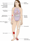
Fig 1
Fig 1
Schematic highlighting the sites and types of polymicrobial diseases commonly located throughout the human body.
Fig 2
Fig 2
Polymicrobial biofilm formation is thought to proceed in several distinct phases. (A) Uncolonized biotic surface (e.g., teeth) lacking any biofilm formation. (B) Deposition of a conditioning layer promotes the adherence of early colonizers that begin the coaggregation cascade. From here, the coaggregative development of the polymicrobial biofilm can occur via two possible methods. (C) Early colonizing bacteria may directly support the binding of late colonizers that then facilitate the attachment of several other microbial species. (D) Specific planktonic intermicrobial interactions can lead to phenotypic changes that support the attachment of preaggregated clusters of cells; however, nonaggregated cells remain unable to attach.
Fig 3
Fig 3
Peptide-nucleic acid fluorescent in situ hybridization (PNA-FISH) microscopy image depicting S. aureus (green) preferentially attached to the hyphal cells of C. albicans (red) during polymicrobial biofilm growth in vitro. (Reprinted from reference with permission of John Wiley & Sons.)
Fig 4
Fig 4
Preinfection of the respiratory epithelial cell line A549 with RSV enhances adherence of P5-fimbriated NTHi (B, black arrow) compared to non-virus-infected control cells (A). (Reprinted from reference .)
Fig 5
Fig 5
In vitro biofilm formation of single-species or polymicrobial inocula composed of NTHi (red) and/or M. catarrhalis (green), assessed over a time course by immunostaining and confocal laser scanning microscopy (CLSM). While single-species biofilms appeared to be composed of smaller aggregates of cells, polymicrobial biofilm growth resulted in larger aggregates of M. catarrhalis interspersed among lawns of NTHi. (Reprinted from reference .)
Fig 6
Fig 6
Scanning electron micrograph comparing in vivo polymicrobial diabetic foot wound growth with in vitro growth obtained by using the Lubbock chronic wound model system. Note the complexity of the adherent microorganism composition growing within close proximity (solid and dashed arrows depict rod- and coccus-shaped bacteria, respectively). “Host” substrata and the elaboration of microbial biofilm matrix also appear similar in both growth systems. (Reprinted from reference with permission of John Wiley & Sons.).
Fig 7
Fig 7
Phase-contrast microscopy image showing the attachment of P. aeruginosa to the hyphal filaments of C. albicans (left, white arrowheads). Epifluorescence microscopy utilizing the Live/Dead staining system (live cells are stained green, while dead or dying cells appear red) demonstrates the ability of P. aeruginosa to specifically kill the hyphae of C. albicans, while the yeast form remains unharmed (right, white arrowheads). (Reprinted from reference with permission of John Wiley & Sons.)
Fig 8
Fig 8
Live/Dead-stained fluorescence microscopy images of in vitro PEG tube surface biofilms grown in chemostats at pH 6.0 (A), pH 5.0 (B), pH 4.0 (C), and pH 3.0 (D). Note that as the pH decreases, the yeast cell mass and pseudohyphal growth increase. Frequently, bacterial cells surrounding these filamentous fungi were stained red, indicating cell death, while nearby microbial community members not in contact with the pseudohyphae appeared healthy (green). (Reprinted from reference .)
Fig 9
Fig 9
Schematic showing the interdependent relationships required for development of human disease. Infection is influenced by microbe-microbe interactions, microbe-host interactions, antimicrobial host defenses, and environmental factors. Significant changes in any of these factors can lead to the development of or predisposition to infection. For example, microbes lacking virulence factors may become apathogenic. Similarly, host immunodeficiencies will encourage infectious processes. It is now becoming increasingly appreciated that intermicrobial interactions and environmental cues also determine infection outcomes such that specific microbial populations under certain conditions may enhance or predict disease progression.
None
None
None
None
None

References

    1. Aas JA, Paster BJ, Stokes LN, Olsen I, Dewhirst FE. 2005. Defining the normal bacterial flora of the oral cavity. J. Clin. Microbiol. 43:5721–5732 - PMC - PubMed
    1. Abramson JS, Wheeler JG. 1994. Virus-induced neutrophil dysfunction: role in the pathogenesis of bacterial infections. Pediatr. Infect. Dis. J. 13:643–652 - PubMed
    1. Alberti KG, Zimmet PZ. 1998. Definition, diagnosis and classification of diabetes mellitus and its complications. Part 1: diagnosis and classification of diabetes mellitus provisional report of a WHO consultation. Diabet. Med. 15:539–553 - PubMed
    1. Al-Fattani MA, Douglas LJ. 2006. Biofilm matrix of Candida albicans and Candida tropicalis: chemical composition and role in drug resistance. J. Med. Microbiol. 55:999–1008 - PubMed
    1. Andreasen C, Carbonetti NH. 2008. Pertussis toxin inhibits early chemokine production to delay neutrophil recruitment in response to Bordetella pertussis respiratory tract infection in mice. Infect. Immun. 76:5139–5148 - PMC - PubMed

Publication types

MeSH terms