

Substratum topography modulates corneal fibroblast to myofibroblast transformation
- PMID: 22232431
- PMCID: PMC3317421
- DOI: 10.1167/iovs.11-7982
Substratum topography modulates corneal fibroblast to myofibroblast transformation
Abstract
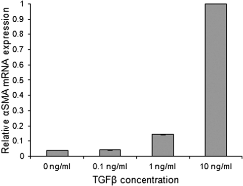
Purpose: The transition of corneal fibroblasts to the myofibroblast phenotype is known to be important in wound healing. The purpose of this study was to determine the effect of topographic cues on TGFβ-induced myofibroblast transformation of corneal cells.
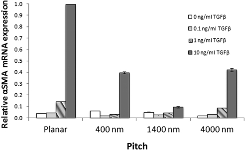
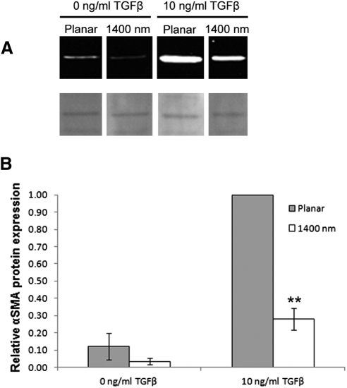
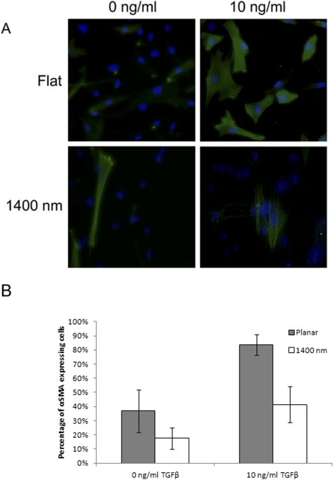
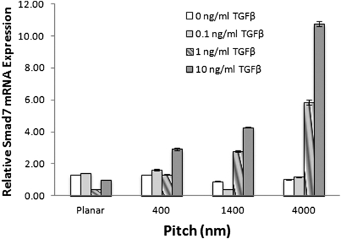
Methods: Rabbit corneal fibroblasts were cultured on nanopatterned surfaces having topographic features of varying sizes. Cells were cultured in media containing TGFβ at concentrations ranging from 0 to 10 ng/mL. RNA and protein were collected from cells cultured on topographically patterned and planar substrates and analyzed for the myofibroblast marker α-smooth muscle actin (αSMA) and Smad7 expression by quantitative real time PCR. Western blot and immunocytochemistry analysis for αSMA were also performed.
Results: Cells grown on patterned surfaces demonstrated significantly reduced levels of αSMA (P < 0.002) compared with planar surfaces when exposed to TGFβ; the greatest reduction was seen on the 1400 nm surface. Smad7 mRNA expression was significantly greater on all patterned surfaces exposed to TGFβ (P < 0.002), whereas cells grown on planar surfaces showed equal or reduced levels of Smad7. Western blot analysis and αSMA immunocytochemical staining demonstrated reduced transition to the myofibroblast phenotype on the 1400 nm surface when compared with cells on a planar surface.
Conclusions: These data demonstrate that nanoscale topographic features modulate TGFβ-induced myofibroblast differentiation and αSMA expression, possibly through upregulation of Smad7. It is therefore proposed that in the wound environment, native nanotopographic cues assist in stabilizing the keratocyte/fibroblast phenotype while pathologic microenvironmental alterations may be permissive for increased myofibroblast differentiation and the development of fibrosis and corneal haze.
Figures
Similar articles
-
Substratum compliance modulates corneal fibroblast to myofibroblast transformation.Invest Ophthalmol Vis Sci. 2013 Aug 28;54(8):5901-7. doi: 10.1167/iovs.12-11575. Invest Ophthalmol Vis Sci. 2013. PMID: 23860754 Free PMC article.
-
Modulation of human corneal stromal cell differentiation by hepatocyte growth factor and substratum compliance.Exp Eye Res. 2018 Nov;176:235-242. doi: 10.1016/j.exer.2018.09.001. Epub 2018 Sep 5. Exp Eye Res. 2018. PMID: 30193807 Free PMC article.
-
Latrunculin B and substratum stiffness regulate corneal fibroblast to myofibroblast transformation.Exp Eye Res. 2018 May;170:101-107. doi: 10.1016/j.exer.2018.02.003. Epub 2018 Feb 6. Exp Eye Res. 2018. PMID: 29421383 Free PMC article.
-
The corneal fibrosis response to epithelial-stromal injury.Exp Eye Res. 2016 Jan;142:110-8. doi: 10.1016/j.exer.2014.09.012. Exp Eye Res. 2016. PMID: 26675407 Free PMC article. Review.
-
Meet the corneal myofibroblast: the role of myofibroblast transformation in corneal wound healing and pathology.Vet Ophthalmol. 2009 Nov-Dec;12 Suppl 1(Suppl 1):25-7. doi: 10.1111/j.1463-5224.2009.00742.x. Vet Ophthalmol. 2009. PMID: 19891648 Free PMC article. Review.
Cited by
-
A high-throughput microfluidic method for fabricating aligned collagen fibrils to study Keratocyte behavior.Biomed Microdevices. 2019 Nov 18;21(4):99. doi: 10.1007/s10544-019-0436-3. Biomed Microdevices. 2019. PMID: 31741114 Free PMC article.
-
Metal Oxide Engineered Nanomaterials Modulate Rabbit Corneal Fibroblast to Myofibroblast Transformation.Transl Vis Sci Technol. 2021 Oct 4;10(12):23. doi: 10.1167/tvst.10.12.23. Transl Vis Sci Technol. 2021. PMID: 34661622 Free PMC article.
-
Biomechanical relationships between the corneal endothelium and Descemet's membrane.Exp Eye Res. 2016 Nov;152:57-70. doi: 10.1016/j.exer.2016.09.004. Epub 2016 Sep 14. Exp Eye Res. 2016. PMID: 27639516 Free PMC article. Review.
-
Junctional adhesion molecule-A regulates vascular endothelial growth factor receptor-2 signaling-dependent mouse corneal wound healing.PLoS One. 2013 May 8;8(5):e63674. doi: 10.1371/journal.pone.0063674. Print 2013. PLoS One. 2013. PMID: 23667656 Free PMC article.
-
Andrographolide Inhibits Corneal Fibroblast to Myofibroblast Differentiation In Vitro.Biomolecules. 2022 Oct 9;12(10):1447. doi: 10.3390/biom12101447. Biomolecules. 2022. PMID: 36291655 Free PMC article.
References
-
- Snyder MC, Bergmanson JP, Doughty MJ. Keratocytes: no more the quiet cells. J Am Optom Assoc. 1998;69:180–187 - PubMed
-
- Jester JV, Petroll WM, Barry PA, Cavanagh HD. Expression of alpha-smooth muscle (alpha-SM) actin during corneal stromal wound healing. Invest Ophthalmol Vis Sci. 1995;36:809–819 - PubMed
-
- Netto MV, Mohan RR, Ambrosio R, Jr, Hutcheon AE, Zieske JD, Wilson SE. Wound healing in the cornea: a review of refractive surgery complications and new prospects for therapy. Cornea. 2005;24:509–522 - PubMed
-
- Saika S, Yamanaka O, Sumioka T, et al. Fibrotic disorders in the eye: targets of gene therapy. Prog Retin Eye Res. 2008;27:177–196 - PubMed
Publication types
MeSH terms
Substances
Grants and funding
LinkOut - more resources
Full Text Sources
Miscellaneous
