. 2012 Apr;81(4):556-66.
doi: 10.1124/mol.111.076125. Epub 2012 Jan 9.

cAMP-specific phosphodiesterases 8A and 8B, essential regulators of Leydig cell steroidogenesis

Affiliations

cAMP-specific phosphodiesterases 8A and 8B, essential regulators of Leydig cell steroidogenesis

Masami Shimizu-Albergine et al. Mol Pharmacol. 2012 Apr.

Abstract

Phosphodiesterase (PDE) 8A and PDE8B are high-affinity, cAMP-specific phosphodiesterases that are highly expressed in Leydig cells. PDE8A is largely associated with mitochondria, whereas PDE8B is broadly distributed in the cytosol. We used a new, PDE8-selective inhibitor, PF-04957325, and genetically ablated PDE8A(-/-), PDE8B(-/-) and PDE8A(-/-)/B(-/-) mice to determine roles for these PDEs in the regulation of testosterone production. PF-04957325 treatment of WT Leydig cells or MA10 cells increased steroid production but had no effect in PDE8A (-/-)/B(-/-) double-knockout cells, confirming the selectivity of the drug. Moreover, under basal conditions, cotreatment with PF-04957325 plus rolipram, a PDE4-selective inhibitor, synergistically potentiated steroid production. These results suggest that the pool(s) of cAMP regulating androgen production are controlled by PDE8s working in conjunction with PDE4. Likewise, PDE8A (-/-)/B(-/-) cells had higher testosterone production than cells from either PDE8A(-/-) or PDE8B(-/-) mice, suggesting that both PDE8s work in concert to regulate steroid production. We further demonstrate that combined inhibition of PDE8s and PDE4 greatly increased PKA activity including phosphorylation of cholesterol-ester hydrolase (CEH)/hormone-sensitive lipase (HSL). CEH/HSL phosphorylation also was increased in PDE8A(-/-)/B(-/-) cells compared with WT cells. Finally, combined inhibition of PDE8s and PDE4 increased the expression of steroidogenic acute regulatory (StAR) protein. Together these findings suggest that both PDE8A and PDE8B play essential roles to maintain low cAMP levels, thereby suppressing resting steroidogenesis by keeping CEH/HSL inactive and StAR protein expression low. They also suggest that in order for PDE inhibitor therapy to be an effective stimulator of steroidogenesis, both PDE8 isozymes and PDE4 need to be simultaneously targeted.


Figures

Fig. 1.
Fig. 1.
Expression of PDE8A and PDE8B in Leydig cells in mouse testis. Sections from PDE8A(−/−), PDE8B(−/−), and WT mouse testes were immunostained with β-galactosidase antibody, as a measure of protein transcribed under the PDE8A or PDE8B promoter. Most of the staining in PDE8A(−/−) and PDE8B(−/−) testes was located in cells containing large nuclei in the interstitial region, which are most likely all Leydig cells. Sperm also expresses both PDE8A and -8B; however, mature sperm do not express this exogenous β-galactosidase, as might be expected.
Fig. 2.
Fig. 2.
Distinct cellular localizations of PDE8A and PDE8B. A, immunostaining for PDE8A and PDE8B was performed in enriched Leydig cells from WT testis as described under Materials and Methods. Immunofluorescence with antibodies specific for PDE8A and PDE8B showed distinct patterns of staining. Signals for PDE8B (red) were broadly distributed in the cytosol. B, signals for PDE8A staining (green) were located in or very near organella, most likely mitochondria, as indicated by the near-perfect overlap with P450scc (red), a known mitochondrial marker protein in these cells. C, immunostaining for PDE8A and PDE8B was performed in MA10 mouse Leydig cell lines. Signals for PDE8A (green) and PDE8B (blue) show a distinct pattern of cellular distribution like that seen in the Leydig cells. D, control cells in which the primary antibodies were omitted showed absence of nonspecific staining. E, immunostaining of PDE8A (green) was entirely overlapped with P450scc (red) as seen in the merged image. All blue staining except in C are nuclei of cells stained with TO-PRO-3. MA10 cells were transfected with either a PDE8A shRNA construct (F) or a negative control (NC) construct (G) and then fixed and processed for immunostaining for PDE8A (red). Depletion of PDE8A by shRNA treatment with PDE8A sequence but not control sequence was seen in the cells expressing the construct marker GFP (green).
Fig. 3.
Fig. 3.
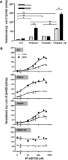
PDE8 inhibition increases resting levels of testosterone production in isolated Leydig cells. Leydig cell-enriched preparations were obtained from WT and PDE8 knockout mice. A, the cells were incubated with or without IBMX (50 μM) for 3 h, and the testosterone content of the media was measured. Results are expressed as amounts of testosterone per Leydig cell-specific 3β-HSD activity in each group. Each data point is expressed as mean ± S.E.M. The data are representative of sets of five experiments. B, the cells were incubated with various doses of PF-04957325 (10–500 nM) with or without IBMX (50 μM) for 3 h. Results are expressed as amount of testosterone per 3β-HSD activity with the curves fitted to a sigmoidal dose-response using Prism. The data are representative sets of three repeated experiments.
Fig. 4.
Fig. 4.
PDE8 inhibition controls testosterone production stimulated by LH in isolated Leydig cells. Leydig cell-enriched preparations were obtained from WT and PDE8 knockout mice. The cells were preincubated with or without PF-04957325 (300 nM) in the presence or absence of IBMX (50 μM) for 30 min and further incubated with different concentrations of LH (30 or 100 pg/ml) for an additional 2.5 h. The testosterone content of the media was measured as described under Materials and Methods. Results are expressed as mean ± S.E.M. of amounts of testosterone per 3β-HSD activity in each preparation. The black bars represent testosterone secreted under no LH stimulation at the resting state. The white bars overlapped with black bars represent the testosterone secreted under stimulation with 30 pg/ml LH. The gray bars overlapped with black bars represent the testosterone secreted under stimulation with 100 pg/ml LH. Statistical analyses were performed by unpaired Student's t test (two-tailed); *, p < 0.05; **, p < 0.01 versus control without PF and IBMX in each group. The data are representative sets of three repeated experiments.
Fig. 5.
Fig. 5.
The effect of PDE8 inhibition on resting levels of progesterone production is augmented by concomitant PDE4 inhibition. A, MA10 cells were incubated with various doses of PF-04957325 (3–1000 nM) for 3 h. The progesterone content of the media was measured as described under Materials and Methods. Results are expressed as amounts of progesterone per protein with the curves fitted to a sigmoidal dose-response using Prism. B, MA10 cells were incubated with PF-04957325 (300 nM) in the presence of IBMX (50 μM) or rolipram (20 μM) for 3 h. The data are representative sets of three repeated experiments. C and D, the cells were preincubated with either vehicle, PF-04957325 (100 nM), IBMX (40 μM), or PF-04957325 plus IBMX for 30 min and incubated with various concentrations of LH (0.1–100 ng/ml) or forskolin (0.1- 50 μM) in the presence of the PDE inhibitors for an additional 2.5 h. Results are expressed as amounts of progesterone per protein with the curves fitted using Prism. The data are representative sets of three repeated experiments.
Fig. 6.
Fig. 6.
One or more PDE4s are the secondary PDEs for regulation of the resting levels of steroid production in Leydig cells. A, Leydig cell-enriched preparations were obtained from WT and PDE8A(−/−)/B(−/−) mice. The cells were incubated with PF-04957325 (300 nM, PDE8 inhibitor), IBMX (50 μM, nonselective PDE inhibitor), rolipram (20 μM, PDE4 inhibitor), SCH51866 (10 μM, PDE1 inhibitor), cilostamide (1 μM, PDE3 inhibitor), Bay 60–7550 (1 μM, PDE2 inhibitor), or sildenafil (100 nM, PDE5 inhibitor) for 3 h, and the content of testosterone in the media was measured. Only PF-04957325 increased the resting level of testosterone production in WT cells, whereas in PDE8A(−/−)/B(−/−) cells, IBMX and rolipram could increase testosterone production. Each data point was expressed as mean ± S.D. Statistical analyses were performed by using unpaired student's t test (two-tailed); **, p < 0.01 versus no inhibitor. The data are representative sets of 3 repeated experiments. B, MA10 cells were incubated with either IBMX (50 μM), rolipram (20 μM), SCH51866 (10 μM), cilostamide (1 μM), Bay 60–7550 (1 μM) or sildenafil (100 nM) in the presence or absence of PF-04957325 (300 nM) for 3 h. Each data point was expressed as mean ± S.D. Statistical analyses were performed by unpaired Student's t test (two-tailed); *, p < 0.05 versus no inhibitor treatment; **, p < 0.01 versus PF-04957325 treatment. The data are representative sets of four repeated experiments.
Fig. 7.
Fig. 7.
PDE8 inhibition increases PKA-dependent phosphorylation and protein expression of components of steroidogenesis. A, MA10 cells were incubated with or without PF-04957325 (concentrations indicated in figure) in the presence or absence of IBMX (50 μM) or rolipram (20 μM) for 3 h. The cell lysates were immunoblotted with anti-phospho-PKA substrate antibody or anti-phospho-HSL (Ser660) antibody. GAPDH was used as a loading control. B, PF-04957325 stimulated a dose-depend phosphorylation of HSL and induction of StAR protein in the presence of IBMX in MA10 cells. Immunoblot analysis of the cell lysates was carried out with anti-phospho-HSL (Ser660), anti-HSL, or anti-StAR antibody. Representative data were shown here. All experiments were repeated at least three times and produced nearly identical results. C, isolated Leydig cell-enriched preparations were obtained from WT, PDE8A(−/−), PDE8B(−/−), and PDE8A(−/−)/B(−/−). The cell lysates were used for immunoblot analysis of phopho-HSL (Ser660) or total HSL. GAPDH was used as a loading control. All immunoblots are representative set of three repeated experiments. D, a quantitative analysis of phospho-HSL content in isolated Leydig cell preparations. Phosphoantibody-specific bands were scanned by densitometry and are represented as a ratio to GAPDH expression. The data are expressed as a fold increase in the ratio of phospho-HSL to GAPDH of WT and represent the mean ± S.D. of three independent experiments. **, p < 0.01 versus WT analyzed by Mann-Whitney test.
Fig. 8.
Fig. 8.
Model of cAMP compartmentalization controlled by PDE8 or PDE4 in regulation of steroidogenesis in Leydig cells. The low Km and Vmax of PDE8A and -8B are depicted as predominant regulators of the resting state of cAMP, thereby keeping steroid production low. PDE8A and PDE4 share, at least in part, some of the same cAMP compartment. PDE4 inhibition alone has very little effect on resting levels of steroid production when PDE8A is still active. Under higher agonist stimulation, the low-Km PDE8s are overwhelmed by the high rate of cAMP synthesis. The higher-Km PDE4s then become the most important regulators at stimulated levels of cAMP synthesis and thereby steroid production.

References

    1. Anthonsen MW, Rönnstrand L, Wernstedt C, Degerman E, Holm C. (1998) Identification of novel phosphorylation sites in hormone-sensitive lipase that are phosphorylated in response to isoproterenol and govern activation properties in vitro. J Biol Chem 273:215–221 - PubMed
    1. Arakane F, King SR, Du Y, Kallen CB, Walsh LP, Watari H, Stocco DM, Strauss JF., 3rd (1997) Phosphorylation of steroidogenic acute regulatory protein (StAR) modulates its steroidogenic activity. J Biol Chem 272:32656–32662 - PubMed
    1. Ascoli M. (1981) Characterization of several clonal lines of cultured Leydig tumor cells: gonadotropin receptors and steroidogenic responses. Endocrinology 108:88–95 - PubMed
    1. Carlucci A, Lignitto L, Feliciello A. (2008) Control of mitochondria dynamics and oxidative metabolism by cAMP, AKAPs and the proteasome. Trends Cell Biol 18:604–613 - PubMed
    1. Conti M, Beavo J. (2007) Biochemistry and physiology of cyclic nucleotide phosphodiesterases: essential components in cyclic nucleotide signaling. Annu Rev Biochem 76:481–511 - PubMed

Publication types

LinkOut - more resources