. 2012 Mar 2;287(10):7427-35.
doi: 10.1074/jbc.M111.324368. Epub 2012 Jan 9.

Non-viral expression of mouse Oct4, Sox2, and Klf4 transcription factors efficiently reprograms tadpole muscle fibers in vivo

Affiliations

Non-viral expression of mouse Oct4, Sox2, and Klf4 transcription factors efficiently reprograms tadpole muscle fibers in vivo

Céline Vivien et al. J Biol Chem. .

Erratum in

  • J Biol Chem. 2012 Jun 22;287(26):22151

Abstract

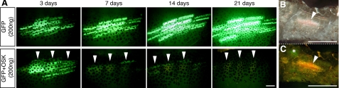
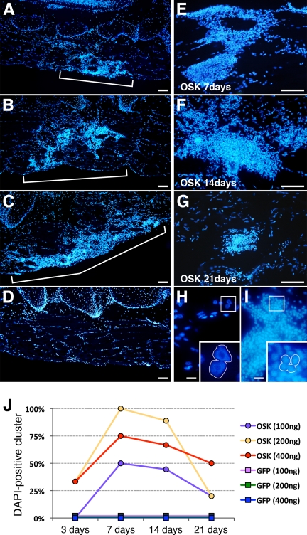
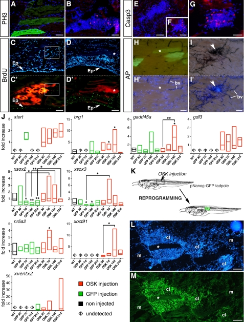
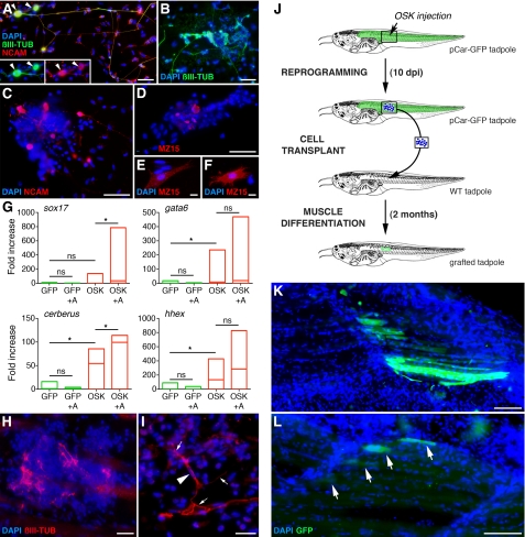
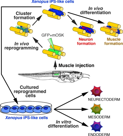
Adult mammalian cells can be reprogrammed into induced pluripotent stem cells (iPSCs) by a limited combination of transcription factors. To date, most current iPSC generation protocols rely on viral vector usage in vitro, using cells removed from their physiological context. Such protocols are hindered by low derivation efficiency and risks associated with genome modifications of reprogrammed cells. Here, we reprogrammed cells in an in vivo context using non-viral somatic transgenesis in Xenopus tadpole tail muscle, a setting that provides long term expression of non-integrated transgenes in vivo. Expression of mouse mOct4, mSox2, and mKlf4 (OSK) led rapidly and reliably to formation of proliferating cell clusters. These clusters displayed the principal hallmarks of pluripotency: alkaline phosphatase activity, up-regulation of key epigenetic and chromatin remodeling markers, and reexpression of endogenous pluripotent markers. Furthermore, these clusters were capable of differentiating into derivatives of the three germ layers in vitro and into neurons and muscle fibers in vivo. As in situ reprogramming occurs along with muscle tissue repair, the data provide a link between these two processes and suggest that they act synergistically. Notably, every OSK injection resulted in cluster formation. We conclude that reprogramming is achievable in an anamniote model and propose that in vivo approaches could provide rapid and efficient alternative for non-viral iPSC production. The work opens new perspectives in basic stem cell research and in the longer term prospect of regenerative medicine protocols development.


Figures

FIGURE 1.
FIGURE 1.
Expression of mouse OSK factors in tadpole muscle leads to GFP loss and cell mass appearance. A, somatic transgenesis in tadpole tail muscle of a GFP-reporter construct ± mouse OSK (200 ng/μl per plasmid). GFP expression was monitored at 3, 7, 14, and 21 dpi, increasing in controls (upper panels) but decreasing at 7 dpi in OSK-injected muscles (lower panels; arrowheads indicate the same myomeric units). Experiments were performed ≥5 times (n >10 for each time point), providing similar results. B, 200-μm-thick slice from OSK muscle at 11 dpi revealing a large, dense cell mass. C, residual GFP-positive fiber (*) near the mass (arrowhead). Scale bars, 500 μm.
FIGURE 2.
FIGURE 2.
Cell cluster formation is dose- and time-dependent. A–D, nuclei, observed by DAPI-staining, are grouped in OSK clusters at 7 dpi (A, 100 ng/μl; B, 200 ng/μl; C, 400 ng/μl for each plasmid), whereas muscle fibers show normal peripheral nuclear organization in GFP-controls (D). E–G, representative clusters surround neighboring muscle fibers at 7, 14, and 21 dpi for the 200 ng/μl condition. H and I, magnifications show large nuclei in control fibers (H), whereas nuclei in OSK clusters are smaller (I). J, kinetics of cluster occurrence for GFP-controls and OSK-injected muscles shown at the 3 concentrations. For each point, independent 4 ≤ n ≤ 9 samples were pooled and the percentage of cluster-containing muscles calculated. Scale bars, 100 μm (A–G), 20 μm (H–I).
FIGURE 3.
FIGURE 3.
Cell clusters are proliferative and express endogenous pluripotency markers. Using PH3-antibody (A and B) and BrdU labeling (C′ and D′), cell proliferation was compared at 14 dpi between GFP (A) or RFP (C and C′) controls and OSK-injected muscles (B, D, and D′). PH3-positive cells (B) and strong BrdU labeling (D′) occurred in OSK clusters, but not in controls (A and C′). Note that BrdU labeling shows similar proliferative status of epidermis (Ep) in both conditions. Apoptosis was followed using active caspase 3 antibody in OSK-transfected muscles at 14 dpi (E and F) and 21 dpi (G), showing an increase of apoptotic cells in clusters. AP activity was strong in OSK-injected muscles at 14 dpi and co-localized with DAPI-positive clusters (I and I′, arrows), whereas GFP-controls (G and G′) showed no AP labeling except in blood vessels (bv). Each labeling was performed ≥3 times. Scale bars, 100 μm (A–E and H–I′); 50 μm (F and G). J, expression of xtert, brg1, and gadd45a (epigenetic and chromatin-remodeling markers) and nr5a2, xoct91, gdf3, xsox3, xsox2, and xventx2 (pluripotency factors) at 3, 7, 14, and 21 dpi in GFP-controls (green boxes) and OSK-transfected muscles (red boxes), using real-time quantitative PCR. For each gene, basal levels in non-injected muscles are indicated (WT, black boxes) and absence of transcript noted as Φ. Samples were from independent experiments, with three muscles pooled per sample (4 ≤ n ≤ 8 samples per group). mRNA levels were expressed as relative to WT except when expression was not detected, then mRNA levels were expressed as relative to the first value observed for OSK. Boxes represent minimum and maximum values around the median. Non-parametric Mann-Whitney U test was used to assess statistical differences versus WT, except when indicated by black bars: **, p < 0.01; *, p < 0.05. K, schematic representation of the protocol used in reprogramming experiment performed with the pNanog-GFP transgenic tadpoles. L and M, GFP immunolabeling of a pNanog-GFP transgenic Xenopus tadpole injected with OSK shows a strong and specific GFP expression in clusters (cl) at 7 dpi, whereas muscle fibers (m and *) are GFP-negative. Scale bars, 100 μm (K–L).
FIGURE 4.
FIGURE 4.
OSK-reprogrammed cells can differentiate into derivates of the three embryonic lineages in vitro and into neurons and muscles in vivo. A–C, neuroectodermal differentiation with FGF-8 + Noggin + retinoic acid (A) or FGF-8 + Noggin (B and C) treatment of 12 dpi OSK-derived cultures, shows tubulin III labeled neurons after 5 days (A and B), and NCAM-positive cells at 3 days (C). D–F, mesodermal differentiation was obtained with low activin treatment of 12 dpi OSK-derived cultures and revealed with a notochord (MZ15) antibody, showing MZ15-positive cells after 3 days (D, activin 5 ng/ml), or individual labeled cells at 3 days (F, activin 50 ng/ml) or 5 days (E, activin 5 ng/ml). G, endodermal differentiation was obtained with high activin (100 ng/ml) + Noggin treatments of 12 dpi OSK-derived cultures and revealed by real-time quantitative PCR using endodermal markers (sox17, gata6, cerberus, and hhex). After 3 days, spontaneous as well as induced endoderm induction was observed in non-treated (OSK) and treated (OSK+A) cultures, but not in 12 dpi GFP-control cultures (GFP and GFP+A). Treatments were performed >6 times. Quantitative PCR data are represented as described in Fig. 3 and mRNA levels expressed as relative to control cultures (GFP): *, p < 0.05; not significant (ns) p > 0.1. H and I, OSK-injected muscles labeled with anti-tubulin III antibody at 14 dpi showed spontaneous neuronal differentiation in cell clusters (H). I, differentiated neuron, with axonal network (arrows) and its cell body (arrowhead indicates nucleus), was obtained following 5 days treatment of 12 dpi OSK-injected tadpoles with FGF-8 + Noggin. J, schematic represents transplant protocol used to follow reprogrammed cell fate reversion in muscle tissue. OSK was injected in a pCar-GFP transgenic tadpole, at 10 dpi the reprogrammed cells were transplanted in a WT tadpole, and the muscle of grafted tadpoles was observed 2 months later. K and L, two examples of grafted tadpoles show the presence of numerous (K) and at a lesser extent (L, arrows) GFP-positive muscle fibers, indicating the reversion of pCar-GFP reprogrammed cells toward a muscle fiber phenotype after transplantation. Scale bars, 50 μm (A–D), 10 μm (E and F), 25 μm (H and I), 100 μm (K and L).
FIGURE 5.
FIGURE 5.
Generation of Xenopus-iPS-like cells in vivo. Model for in vivo non-viral induction of Xenopus reprogrammed cells is shown. Following co-injection of mOSK with a GFP-reporter in tadpole tail (mOSK+GFP), GFP-transfected fibers (in green) dedifferentiate into proliferative undifferentiated Xenopus iPS-like cells (in blue). Reprogrammed cells of mesodermal origin possess an increased developmental potential, differentiating in vitro toward derivatives of the three embryonic lineages and into neurons and muscle fibers in vivo.

References

    1. Takahashi K., Yamanaka S. (2006) Induction of pluripotent stem cells from mouse embryonic and adult fibroblast cultures by defined factors. Cell 126, 663–676 - PubMed
    1. Ho R., Chronis C., Plath K. (2011) Mechanistic insights into reprogramming to induced pluripotency. J. Cell. Physiol. 226, 868–878 - PMC - PubMed
    1. Tan K. Y., Eminli S., Hettmer S., Hochedlinger K., Wagers A. J. (2011) Efficient generation of iPS cells from skeletal muscle stem cells. PLoS One 6, e26406. - PMC - PubMed
    1. Jaenisch R., Young R. (2008) Stem cells, the molecular circuitry of pluripotency and nuclear reprogramming. Cell 132, 567–582 - PMC - PubMed
    1. Stadtfeld M., Hochedlinger K. (2010) Induced pluripotency: history, mechanisms, and applications. Genes Dev. 24, 2239–2263 - PMC - PubMed

Publication types

MeSH terms

LinkOut - more resources