. 2012 Mar 2;287(10):7265-78.
doi: 10.1074/jbc.M111.287599. Epub 2012 Jan 9.

Flotillin-1/reggie-2 protein plays dual role in activation of receptor-tyrosine kinase/mitogen-activated protein kinase signaling

Affiliations

Flotillin-1/reggie-2 protein plays dual role in activation of receptor-tyrosine kinase/mitogen-activated protein kinase signaling

Monia Amaddii et al. J Biol Chem. .

Abstract

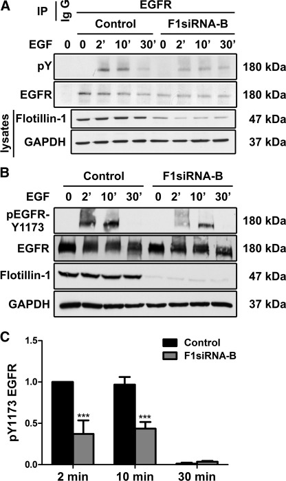
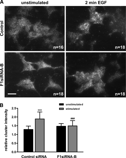
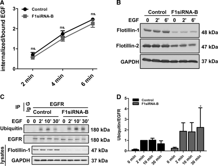
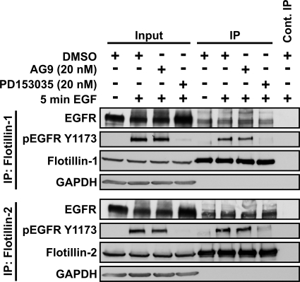
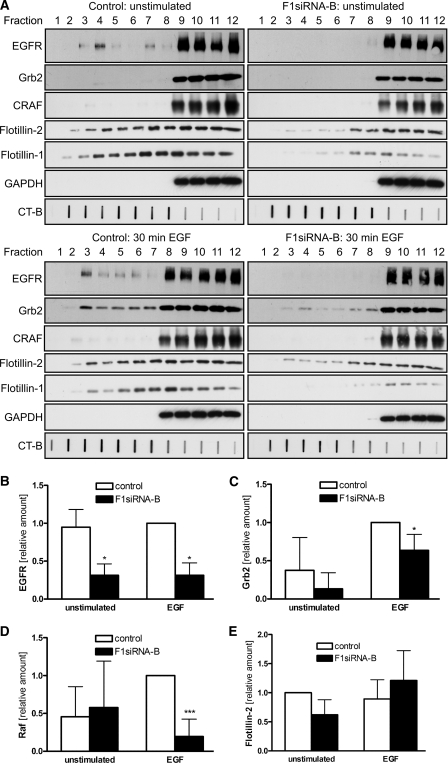
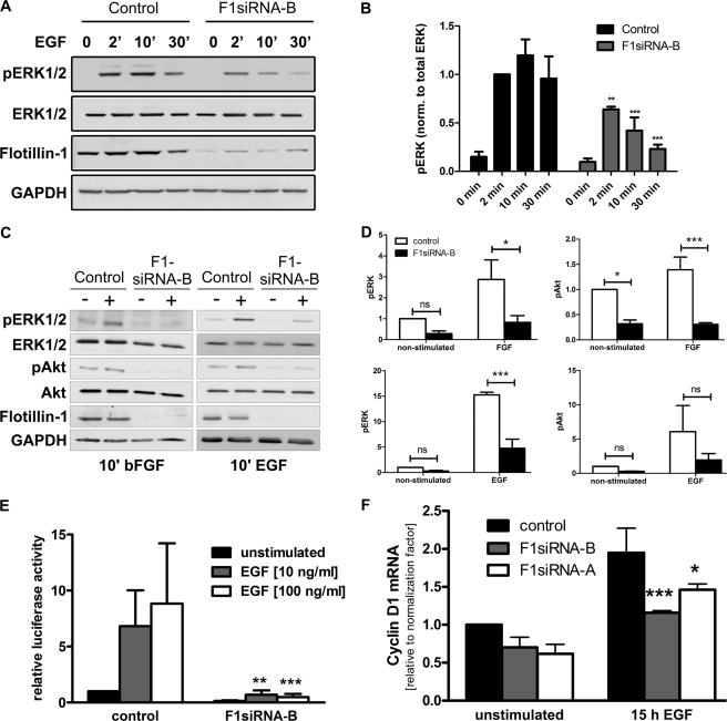
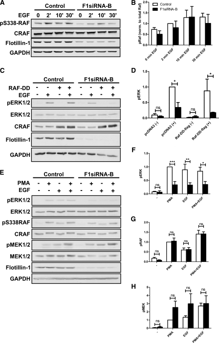
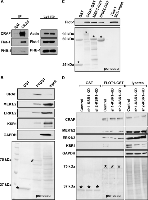
Our previous work has shown that the membrane microdomain-associated flotillin proteins are potentially involved in epidermal growth factor (EGF) receptor signaling. Here we show that knockdown of flotillin-1/reggie-2 results in reduced EGF-induced phosphorylation of specific tyrosines in the EGF receptor (EGFR) and in inefficient activation of the downstream mitogen-activated protein (MAP) kinase and Akt signaling. Although flotillin-1 has been implicated in endocytosis, its depletion affects neither the endocytosis nor the ubiquitination of the EGFR. However, EGF-induced clustering of EGFR at the cell surface is altered in cells lacking flotillin-1. Furthermore, we show that flotillins form molecular complexes with EGFR in an EGF/EGFR kinase-independent manner. However, knockdown of flotillin-1 appears to affect the activation of the downstream MAP kinase signaling more directly. We here show that flotillin-1 forms a complex with CRAF, MEK1, ERK, and KSR1 (kinase suppressor of RAS) and that flotillin-1 knockdown leads to a direct inactivation of ERK1/2. Thus, flotillin-1 plays a direct role during both the early phase (activation of the receptor) and late (activation of MAP kinases) phase of growth factor signaling. Our results here unveil a novel role for flotillin-1 as a scaffolding factor in the regulation of classical MAP kinase signaling. Furthermore, our results imply that other receptor-tyrosine kinases may also rely on flotillin-1 upon activation, thus suggesting a general role for flotillin-1 as a novel factor in receptor-tyrosine kinase/MAP kinase signaling.


Figures

FIGURE 1.
FIGURE 1.
Knockdown of flot-1 results in reduced Tyr phosphorylation of EGFR. Flot-1 was depleted by means of siRNAs. The cells were subsequently starved and stimulated or not with EGF for 2, 10, or 30 min, and EGFR was immunoprecipitated (IP). Precipitated proteins were separated by SDS-PAGE and probed with the indicated antibodies. The cell lysates were analyzed with the monoclonal antibody against flot-1 to verify its depletion, and GAPDH was used as an equal loading control. Effect of flotillin knockdown on total tyrosine phosphorylation (pY; A) and Tyr-1173 (B) was measured. C, the bar graph show a densitometric quantification of the phosphorylation of Tyr-1173. The signals of phosphorylated Tyr-1173 were normalized to the amount of total EGFR. Bars represent the mean ± S.D. of at least three individual experiments. ***, p < 0.001.
FIGURE 2.
FIGURE 2.
EGF-induced clustering of EGFR at the cell surface is impaired by flot-1 knockdown. Flot-1 was knocked down in HeLa cells by siRNA. Knockdown and control cells were starved and stimulated or not for 2 min with EGF. The cells were fixed and immunostained for EGF receptor. Cell surface associated EGFR was detected by total internal reflection microscopy. The images (A) were analyzed for the mean EGFR cluster intensity. B, shown is quantification of the EGFR cluster intensity upon EGF stimulation in control and F1-KD cells (n = 3). ***, p < 0.001 compared to unstimulated control; ###, p > 0.001 compared to stimulated control.
FIGURE 3.
FIGURE 3.
Flot-1 depletion does not inhibit EGFR endocytosis and ubiquitination. F1-KD and control cells were incubated with 125I-EGF (1.5 ng/ml) for the indicated time points, and the internalized versus cell surface-bound EGFs were measured. The data are a summary of three independent experiments, each with triplicate samples for each time point. A, the graph shows the ratio of internalized to surface bound EGF as a function of endocytosis time, including the S.D. The difference between control cells and F1-KD cells was not significant (ns.). B, F1-KD efficiency in the cells used for the experiment is shown. C, Flot-1 was depleted by means of RNAi in HeLa cells that were subsequently starved and stimulated or not with EGF for 2, 10, or 30 min. EGFR was immunoprecipitated (IP) from the cells, and the precipitates were probed with an anti-ubiquitin antibody. IgG, an isotype matched control antibody was used to show the specificity. D, quantification of EGFR ubiquitination from four individual experiments is shown. Ubiquitin signals were normalized to total EGFR. *, p < 0.05.
FIGURE 4.
FIGURE 4.
EGFR co-immunoprecipitates with flotillins. HeLa cells (serum-grown, unstimulated, or EGF stimulated) were lysed and immunoprecipitated with antibodies against flot-2. Co-immunoprecipitation of EGFR with flotillins was detected in all samples. After 5 min of EGF, a precipitated band (185 kDa) migrated slightly higher than EGFR (180 kDa) in unstimulated samples and corresponded to the Tyr-1173-phosphorylated EGFR, as evidenced by detection with an anti-Tyr(P)-1173 antibody. Flot-1 (47 kDa) coprecipitated with flot-2 (48 kDa) in all samples, whereas no precipitation of EGFR or flotillins was detected in the control immunoprecipitation carried out with an anti-myc antibody (Control IP). Equal loading was verified with GAPDH (37 kDa).
FIGURE 5.
FIGURE 5.
Co-precipitation of EGFR and flotillins is not dependent on EGFR kinase activity. Starved HeLa cells were treated with an EGFR kinase inhibitor PD153035, non-inhibiting analog AG9 (each 20 nm), or DMSO and then stimulated for 5 min with EGF. Immunoprecipitation (IP) was performed with anti-flot-1 or flot-2 antibodies. Co-precipitation of EGFR with both flotillins was detected also in samples treated with PD153035, and Tyr-1173-phosphorylated EGFR was coprecipitated in the samples not treated with the inhibitor but treated with EGF. For the molecular masses of the proteins, please refer to the legend of Fig. 4.
FIGURE 6.
FIGURE 6.
EGFR and Grb2 do not translocate into light fractions in F1-KD cells. Flot-1 was knocked down in HeLa cells by siRNA. The cells were starved and stimulated with 100 ng/ml EGF for 30 min on ice or left untreated. A, rafts were isolated from the cells using a detergent-free method. Fractions were collected from the top (fraction 1 = the lightest fraction), run on SDS-PAGE, and analyzed by Western blotting. The localization of EGFR (180 kDa), Grb2 (25 kDa), CRAF (74 kDa), flotillins (47/48 kDa), and GAPDH (37 kDa) was studied. The raft marker GM1 was detected with HRP-coupled cholera toxin in a slot blot. Quantification of the relative distribution in the light fractions: EGFR (B); Grb2 (C); CRAF (D); flotillin-2 (E) (summary of four independent experiments). CT-B , cholera toxin B subunit. *, p < 0.05; ***, p < 0.001.
FIGURE 7.
FIGURE 7.
Activation of MAP kinase pathway is reduced after flot-1 knockdown. Flot-1 was knocked down in HeLa cells. The cells were starved and stimulated or not with EGF for 2, 10, or 30 min. Cell lysates were separated by SDS-PAGE and probed with the indicated antibodies. GAPDH was used as an equal loading control. A, knockdown of flot-1 resulted in severe reduction of activation of the MAP kinases ERK1/2 (41/42 kDa). B, densitometric quantification of the pERK1/2 (42/42 kDa) is shown. The values of phosphorylated ERK1/2 were normalized to total ERK. C, in F1-KD cells, activation of ERK1/2 and Akt were measured after a 10-min treatment with bFGF or EGF. D, shown is quantification of the results in C. ns, not significant. E, transcriptional activation after EGF stimulation was measured using a serum-responsive element-based luciferase reporter gene assay. Transcriptional activation in F1-KD cells was severely impaired irrespective of the EGF dose used. F, quantitative real-time PCR was used to measure the mRNA for cyclin D, which was less induced after EGF stimulation of F1-KD cells. B and D–F, bars represent the means of at least three individual experiments with standard deviation. **, p < 0.01; ***, p < 0.001.
FIGURE 8.
FIGURE 8.
Forced activation of CRAF does not result in improved ERK phosphorylation. A, phosphorylation of CRAF on Ser-338 was studied after EGF stimulation in F1-KD and control cells. B, quantification of the phosphorylation of CRAF p338 after normalization to total CRAF (n = 3). C, transfection of F1-KD cells with an active CRAF mutant (RAF-DD) was not able to rescue the reduced phosphorylation of ERK1/2. D, quantification of the data in C (n = 3). E, stimulation of the cells with PMA did not result in improved ERK1/2 phosphorylation in F1-KD cells. F–H, shown is quantification of the phosphorylation data shown in E. F = pERK. G = p338 CRAF. H = pMEK (n = 3). ns, not significant. *, p < 0.05; **, p < 0.01; ***, p < 0.001.
FIGURE 9.
FIGURE 9.
Flotillin-1 functions as a MAP kinase scaffold. A, CRAF was immunoprecipitated (IP) from HeLa cells, and the immunoprecipitates were probed for flot-1 (47 kDa) and prohibitin-1 (PHB-1; 30 kDa), which both coprecipitated with CRAF. B, in flotillin-1-GST (75 kDa) pulldown assays, CRAF (74 kDa), MEK1/2 (45/47 kDa), ERK1/2 (42 kDa), and KSR1 (110 kDa) interacted with flot-1. C, purified recombinant flot-1 directly interacted with GST-tagged CRAF, MEK1, and ERK2. D, KSR1 was knocked down with two different shRNAs in HeLa cells. Pulldown experiments with flotillin-1-GST show that CRAF, MEK1/2, and ERK1/2 are capable of binding to flot-1 in the absence of KSR1. In addition, KSR1 was also found in flot-1 pulldown assays from control cells. B–D, Ponceau staining was used to visualize the fusion proteins (marked by an asterisk) on the blot membranes.

Similar articles

Cited by

References

    1. Levkowitz G., Waterman H., Zamir E., Kam Z., Oved S., Langdon W. Y., Beguinot L., Geiger B., Yarden Y. (1998) c-Cbl/Sli-1 regulates endocytic sorting and ubiquitination of the epidermal growth factor receptor. Genes Dev. 12, 3663–3674 - PMC - PubMed
    1. Carpentier J. L., Gorden P., Anderson R. G., Goldstein J. L., Brown M. S., Cohen S., Orci L. (1982) Co-localization of 125I-epidermal growth factor and ferritin low density lipoprotein in coated pits. A quantitative electron microscopic study in normal and mutant human fibroblasts. J. Cell Biol. 95, 73–77 - PMC - PubMed
    1. Gorden P., Carpentier J. L., Cohen S., Orci L. (1978) Epidermal growth factor. Morphological demonstration of binding, internalization, and lysosomal association in human fibroblasts. Proc. Natl. Acad. Sci. U.S.A. 75, 5025–5029 - PMC - PubMed
    1. Hanover J. A., Willingham M. C., Pastan I. (1984) Kinetics of transit of transferrin and epidermal growth factor through clathrin-coated membranes. Cell 39, 283–293 - PubMed
    1. Waugh M. G., Lawson D., Hsuan J. J. (1999) Epidermal growth factor receptor activation is localized within low buoyant density, non-caveolar membrane domains. Biochem. J. 337, 591–597 - PMC - PubMed

Publication types

MeSH terms