Angiotropic metastatic malignant melanoma in a canine mammary gland
- PMID: 22232646
- PMCID: PMC3251768
- DOI: 10.5625/lar.2011.27.4.353
Angiotropic metastatic malignant melanoma in a canine mammary gland
Abstract
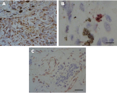
An eleven-year-old spayed female Yorkshire Terrier presented with a sublumbar mass and upon ultrasonographic examination, was revealed to have a mammary gland tumor. Black to reddish colored masses, located in the visceral peritoneum of the sublumbar region was observed on laparotomy with masectomy of the right side. In the laparotomy, we observed reddish masses multifocally located in the serosal membrane of the large intestine. Histopathologic examination of the intestinal and abdominal mass showed highly invasiveness into the muscle and metastasis of melanocytic tumor cells through the blood vessels. The mammary glands showed abnormal hyperplasia of melanocytes, destruction of the normal glands by tumor cells and infiltration of some lymphocytes in the pool of melanocytic cells. We have identified a malignant melanoma containing an angiotumoral complex in which tumor cells occupied a pericytic location along the microvessels with intravasation determined by immunohistochemistry for S100 protein and protein kinase C-α. Histologic findings in this dog lead to a diagnosis of an angiotropic metastatic malignant melanoma.
Keywords: Canine; S100 protein; malignant melanoma; mammary gland; metastasis; protein kinase C-α.
Figures

Similar articles
-
Pericytic-like angiotropism of glioma and melanoma cells.Am J Dermatopathol. 2002 Dec;24(6):473-8. doi: 10.1097/00000372-200212000-00003. Am J Dermatopathol. 2002. PMID: 12454598
-
Metastatic melanoma from an unknown primary site presenting as skin-colored nodules and multiple visceral involvement.Skinmed. 2012 Nov-Dec;10(6):396-9. Skinmed. 2012. PMID: 23346671
-
[A case of malignant melanoma metastasis to the mammary gland].Gan To Kagaku Ryoho. 2014 Nov;41(12):1939-41. Gan To Kagaku Ryoho. 2014. PMID: 25731382 Japanese.
-
Role of In Vivo Reflectance Confocal Microscopy in the Analysis of Melanocytic Lesions.Acta Dermatovenerol Croat. 2018 Apr;26(1):64-67. Acta Dermatovenerol Croat. 2018. PMID: 29782304 Review.
-
Angiotropic melanoma and extravascular migratory metastasis: a review.Adv Anat Pathol. 2007 May;14(3):195-201. doi: 10.1097/PAP.0b013e31805048d9. Adv Anat Pathol. 2007. PMID: 17452816 Review.
References
-
- Bostock DE. Prognosis after surgical excision of canine melanomas. Vet Pathol. 1979;16(1):32–40. - PubMed
-
- Harvey HJ, MacEwen G, Braun D, Patnaik AK, Withrow SJ, Jongeward S. Prognostic criteria for dogs with oral melanoma. J Am Vet Med Assoc. 1981;178(6):580–582. - PubMed
-
- MacEwen EG, Patnaik AK, Harvey HJ, Hayers AA, Matus R. Canine oral melanoma: comparison of surgery versus surgery plus Corynebacterium parvum. Cancer Invest. 1986;4(5):397–402. - PubMed
-
- Ramos-Vara JA, Beissenherz ME, Miller MA, Johnson GC, Pace LW, Fard A, Kottler SJ. Reprospective study of 338 canine oral melanomas with clinical, histologic, and immunohistichemical review of 129 cases. Vet Pathol. 2000;37(6):597–608. - PubMed
-
- Smith SH, Goldschmidt MH, McManus PM. A comparative review of melanocytic neoplasms. Vet Pathol. 2002;39(6):651–678. - PubMed