

Structural basis for membrane targeting by the MVB12-associated β-prism domain of the human ESCRT-I MVB12 subunit
- PMID: 22232651
- PMCID: PMC3277574
- DOI: 10.1073/pnas.1117597109
Structural basis for membrane targeting by the MVB12-associated β-prism domain of the human ESCRT-I MVB12 subunit
Abstract
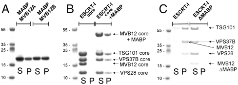
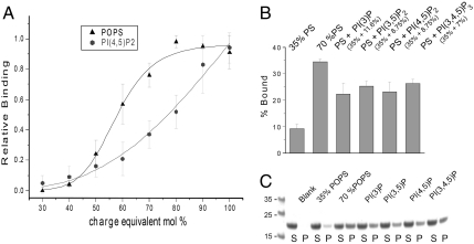
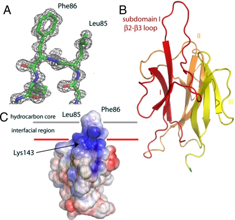
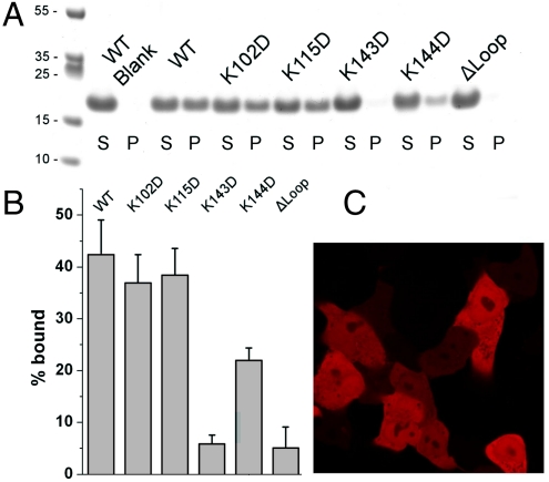
MVB12-associated β-prism (MABP) domains are predicted to occur in a diverse set of membrane-associated bacterial and eukaryotic proteins, but their existence, structure, and biochemical properties have not been characterized experimentally. Here, we find that the MABP domains of the MVB12A and B subunits of ESCRT-I are functional modules that bind in vitro to liposomes containing acidic lipids depending on negative charge density. The MABP domain is capable of autonomously localizing to subcellular puncta and to the plasma membrane. The 1.3-Å atomic resolution crystal structure of the MVB12B MABP domain reveals a β-prism fold, a hydrophobic membrane-anchoring loop, and an electropositive phosphoinositide-binding patch. The basic patch is open, which explains how it senses negative charge density but lacks stereoselectivity. These observations show how ESCRT-I could act as a coincidence detector for acidic phospholipids and protein ligands, enabling it to function both in protein transport at endosomes and in cytokinesis and viral budding at the plasma membrane.
Conflict of interest statement
The authors declare no conflict of interest.
Figures

Comment in
-
Lipid targeting domain with dual-membrane specificity that expands the diversity of intracellular targeting reactions.Proc Natl Acad Sci U S A. 2012 Feb 7;109(6):1816-7. doi: 10.1073/pnas.1120856109. Epub 2012 Jan 26. Proc Natl Acad Sci U S A. 2012. PMID: 22308463 Free PMC article. No abstract available.
References
-
- Hurley JH, Meyer T. Subcellular targeting by membrane lipids. Curr Opin Cell Biol. 2001;13:146–152. - PubMed
-
- Cho WH, Stahelin RV. Membrane-protein interactions in cell signaling and membrane trafficking. Annu Rev Biophys Biomol Struct. 2005;34:119–151. - PubMed
-
- Lemmon MA. Membrane recognition by phospholipid-binding domains. Nat Rev Mol Cell Biol. 2008;9:99–111. - PubMed
Publication types
MeSH terms
Substances
Associated data
- Actions
Grants and funding
LinkOut - more resources
Full Text Sources
Molecular Biology Databases
