

Uric acid stones in the urinary bladder of aryl hydrocarbon receptor (AhR) knockout mice
- PMID: 22232670
- PMCID: PMC3268287
- DOI: 10.1073/pnas.1120581109
Uric acid stones in the urinary bladder of aryl hydrocarbon receptor (AhR) knockout mice
Abstract
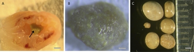
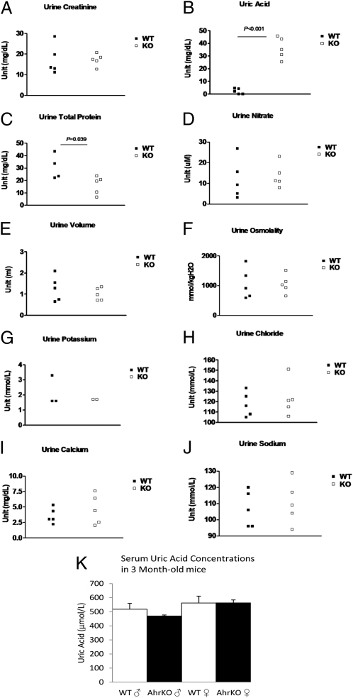
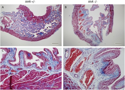
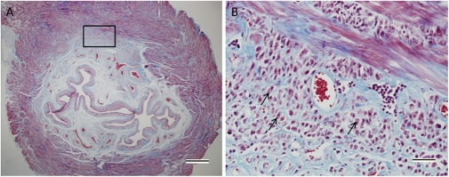
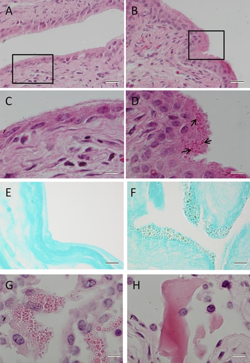
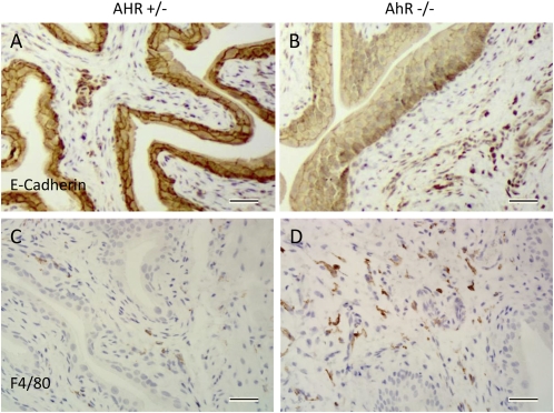
The aryl hydrocarbon receptor (AhR) knockout mice raised in the laboratory of Fujii-Kuriyama have been under investigation for several years because of the presence in their urinary bladder of large, yellowish stones. The stones are composed of uric acid and become apparent in the bladders as tiny stones when mice are 10 wk of age. By the time the mice are 6 mo of age, there are usually two or three stones with diameters of 3-4 mm. The urate concentration in the serum was normal but in the urine the concentration was 40-50 mg/dL, which is 10 times higher than that in the WT littermates. There were no apparent histological pathologies in the kidney or joints and the levels of enzymes involved in elimination of purines were normal. The source of the uric acid was therefore judged to be from degradation of nucleic acids due to a high turnover of cells in the bladder itself. The bladder was fibrotic and the luminal side of the bladder epithelium was filled with eosinophilic granules. There was loss of E-cadherin between some epithelial cells, with an enlarged submucosal area filled with immune cells and sometimes invading epithelial cells. We hypothesize that in the absence of AhR there is loss of detoxifying enzymes, which leads to accumulation of unconjugated cytotoxins and carcinogens in the bladder. The presence of bladder toxins may have led to the increased apoptosis and inflammation as well as invasion of epithelial cells in the bladders of older mice.
Conflict of interest statement
The authors declare no conflict of interest.
Figures

Comment in
-
Re: Uric acid stones in the urinary bladder of aryl hydrocarbon receptor (AhR) knockout mice.J Urol. 2012 Sep;188(3):1042. doi: 10.1016/j.juro.2012.05.040. Epub 2012 Jul 21. J Urol. 2012. PMID: 22883806 No abstract available.
Similar articles
-
Knockout of the aryl hydrocarbon receptor results in distinct hepatic and renal phenotypes in rats and mice.Toxicol Appl Pharmacol. 2013 Oct 15;272(2):503-18. doi: 10.1016/j.taap.2013.06.024. Epub 2013 Jul 13. Toxicol Appl Pharmacol. 2013. PMID: 23859880
-
Local factors compared with systemic factors in the formation of bladder uric acid stones.Urol Int. 2009;82(1):48-52. doi: 10.1159/000176025. Epub 2009 Jan 20. Urol Int. 2009. PMID: 19172097
-
Uric acid bladder stones in congenital cyanotic heart disease.Lancet. 2016 Oct 15;388(10054):1921. doi: 10.1016/S0140-6736(16)00347-0. Epub 2016 May 5. Lancet. 2016. PMID: 27156932 No abstract available.
-
The induction of bladder stones by terephthalic acid, dimethyl terephthalate, and melamine (2,4,6-triamino-s-triazine) and its relevance to risk assessment.Regul Toxicol Pharmacol. 1985 Sep;5(3):294-313. doi: 10.1016/0273-2300(85)90044-3. Regul Toxicol Pharmacol. 1985. PMID: 3903881 Review.
-
Investigations of rodent urinary bladder carcinogens: collection, processing, and evaluation of urine and bladders.Toxicol Pathol. 2007 Apr;35(3):337-47. doi: 10.1080/01926230701197115. Toxicol Pathol. 2007. PMID: 17455081 Review.
Cited by
-
The Aryl Hydrocarbon Receptor (AhR) in the Aging Process: Another Puzzling Role for This Highly Conserved Transcription Factor.Front Physiol. 2020 Jan 14;10:1561. doi: 10.3389/fphys.2019.01561. eCollection 2019. Front Physiol. 2020. PMID: 32009975 Free PMC article. Review.
-
Ah receptor ligands and their impacts on gut resilience: structure-activity effects.Crit Rev Toxicol. 2020 Jul;50(6):463-473. doi: 10.1080/10408444.2020.1773759. Epub 2020 Jun 29. Crit Rev Toxicol. 2020. PMID: 32597352 Free PMC article. Review.
-
The Aryl Hydrocarbon Receptor: A Key Bridging Molecule of External and Internal Chemical Signals.Environ Sci Technol. 2015 Aug 18;49(16):9518-31. doi: 10.1021/acs.est.5b00385. Epub 2015 Aug 10. Environ Sci Technol. 2015. PMID: 26079192 Free PMC article. Review.
-
Aryl Hydrocarbon Receptor (AHR) Ligands as Selective AHR Modulators (SAhRMs).Int J Mol Sci. 2020 Sep 11;21(18):6654. doi: 10.3390/ijms21186654. Int J Mol Sci. 2020. PMID: 32932962 Free PMC article. Review.
-
Aryl Hydrocarbon Receptor (AhR) Ligands as Selective AhR Modulators: Genomic Studies.Curr Opin Toxicol. 2018 Oct-Dec;11-12:10-20. doi: 10.1016/j.cotox.2018.11.005. Epub 2018 Nov 22. Curr Opin Toxicol. 2018. PMID: 31453421 Free PMC article.
References
-
- Nebert DW, Dalton TP, Okey AB, Gonzalez FJ. Role of aryl hydrocarbon receptor-mediated induction of the CYP1 enzymes in environmental toxicity and cancer. J Biol Chem. 2004;279:23847–23850. - PubMed
-
- Sugihara K, et al. Aryl hydrocarbon receptor (AhR)-mediated induction of xanthine oxidase/xanthine dehydrogenase activity by 2,3,7,8-tetrachlorodibenzo-p-dioxin. Biochem Biophys Res Commun. 2001;281:1093–1099. - PubMed
-
- Lin PH, Lin CH, Huang CC, Chuang MC, Lin P. 2,3,7,8-Tetrachlorodibenzo-p-dioxin (TCDD) induces oxidative stress, DNA strand breaks, and poly(ADP-ribose) polymerase-1 activation in human breast carcinoma cell lines. Toxicol Lett. 2007;172:146–158. - PubMed
-
- Muralidhara Matsumura F, Blankenship A. 2,3,7,8-Tetrachlorodibenzo-p-dioxin (TCDD)-induced reduction of adenosine deaminase activity in vivo and in vitro. J Biochem Toxicol. 1994;9:249–259. - PubMed
-
- Moriwaki Y, Yamamoto T, Higashino K. Enzymes involved in purine metabolism—a review of histochemical localization and functional implications. Histol Histopathol. 1999;14:1321–1340. - PubMed
Publication types
MeSH terms
Substances
LinkOut - more resources
Full Text Sources
Molecular Biology Databases
