. 2012 Jan 24;109(4):1293-8.
doi: 10.1073/pnas.1119658109. Epub 2012 Jan 9.

Hypoxic regulation of the cerebral microcirculation is mediated by a carbon monoxide-sensitive hydrogen sulfide pathway

Affiliations

Hypoxic regulation of the cerebral microcirculation is mediated by a carbon monoxide-sensitive hydrogen sulfide pathway

Takayuki Morikawa et al. Proc Natl Acad Sci U S A. .

Abstract

Enhancement of cerebral blood flow by hypoxia is critical for brain function, but signaling systems underlying its regulation have been unclear. We report a pathway mediating hypoxia-induced cerebral vasodilation in studies monitoring vascular disposition in cerebellar slices and in intact mouse brains using two-photon intravital laser scanning microscopy. In this cascade, hypoxia elicits cerebral vasodilation via the coordinate actions of H(2)S formed by cystathionine β-synthase (CBS) and CO generated by heme oxygenase (HO)-2. Hypoxia diminishes CO generation by HO-2, an oxygen sensor. The constitutive CO physiologically inhibits CBS, and hypoxia leads to increased levels of H(2)S that mediate the vasodilation of precapillary arterioles. Mice with targeted deletion of HO-2 or CBS display impaired vascular responses to hypoxia. Thus, in intact adult brain cerebral cortex of HO-2-null mice, imaging mass spectrometry reveals an impaired ability to maintain ATP levels on hypoxia.


Conflict of interest statement

The authors declare no conflict of interest.

Figures

Fig. 1.
Fig. 1.
Immunohistochemical localization of HO-2 and CBS in the neurovascular unit of neonatal mouse cerebellar cortex. (A) HO-2, a CO-producing enzyme, is abundantly expressed in neurons. Note the strong labeling of Purkinje cells (asterisks). (B–D) HO-2 surrounds microvessels. HO-2+ cells along the vessel wall in B are endothelial, not pericytic, because nuclei of cells positive for NG2 (a pericytic marker) stained with TO-PRO-3 (TOPRO, a nucleic acid stain), are completely devoid of CD31 (endothelial marker) labeling in C. In D, the arteriolar wall is surrounded by NG2+ pericytes, important contractile cells within the neurovascular unit. (E–H) CBS, an H2S-producing enzyme, is concentrated at the ascending processes of Bergmann glia (arrow in E and F) and radial processes of astrocytes (arrowheads in E and G), as evidenced by the colocalization with GFAP, an established marker of glial cells. The astrocytic endfeet in contact with the vessel wall are CBS-positive in H. ml, molecular layer; Pl, Purkinje cell layer; gl, granular layer; e, endothelium; p, pericyte. (I) Schematic depiction of localization of HO-2 and CBS in the neurovascular unit.
Fig. 2.
Fig. 2.
Pharmacologic inhibition of endogenous CO production evokes a CBS-dependent arteriolar vasodilation in neonatal mouse cerebellar slices. (A) A typical change in arteriolar diameter of cerebellar slices in response to ZnPP (1 μM), an HO inhibitor. (B) Time course of arteriolar dilation during superfusion of the potent HO inhibitors ZnPP (1 μM) and CrMP (1 μM). (C) Summary of changes in arteriolar diameter at 60 min after superfusion of various reagents. Vasodilation induced by HO inhibition is reversed by tricarbonyldichlororuthenium(II) dimer, [Ru(CO)3Cl2]2, a CO-releasing molecule (CORM-2; 100 μM), indicating that CO acts as a tonic vasoconstrictor. The vasodilatory response of ZnPP does not occur in CBS-null slices. Glib, glibenclimide, an inhibitor of KATP channels. *P < 0.05 compared with the vehicle-treated control; P < 0.05 compared with the ZnPP-treated WT mice; P < 0.05 compared with the ZnPP-treated WT mice. Values are mean ± SEM.
Fig. 3.
Fig. 3.
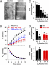
The HO-2/CO and CBS/H2S pathways mediate hypoxia-induced arteriolar vasodilation. (A) Hypoxia-induced arteriolar vasodilation of an arteriole in a cerebellar slice. Dashed lines indicate the previous position of the vessel wall. (B) Lowering the partial pressure of O2 (PO2) in the superfusates is followed by robust arteriolar dilation in WT mice. Deletion of either HO-2 or CBS causes a significant reduction in the extent of hypoxia-induced arteriolar vasodilation. *P < 0.05 compared with WT. (C) Lowering the O2 concentration gradually decreases CO production from cerebellar slices. *P < 0.05 compared with 100% O2. (D) The O2-dependent reduction in CO production is abolished in HO-2-null mice. The CO concentration in HO-2-null mice at 10% O2 is approximately half that in WT mice. *P < 0.05 compared with WT at 10% O2. (E) Endogenous H2S concentration in neonatal brain (P12) measured by the reaction of monobromobimane, an electrophilic reagent, with HS to form sulfide dibimane (SDB). Lowering the O2 concentration elevates the endogenous H2S concentration. Hypoxia-induced elevation of H2S concentrations is abolished in HO-2-null mice. *P < 0.05 compared with WT at 21% O2.
Fig. 4.
Fig. 4.
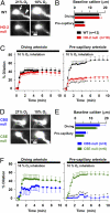
Hypoxia-induced vasodilation of precapillary arterioles in vivo is attenuated in HO-2-null mice and abolished in CBS-null mice. (A) Vasodilatory responses of diving (arrow) and precapillary arterioles (arrowhead) imaged in vivo in live mouse cerebral cortex through the thinned skull at a depth of 50–90 μm using two-photon laser scanning microscopy. Qdot655 was injected i.v. to outline the vasculature. (B) Baseline diameter of arterioles. (C) Inhalation of 10% O2 evokes rapid vasodilation of both diving and precapillary arterioles. HO-2 deletion causes a marked attenuation in the vasodilatory response of precapillary arterioles, but not of diving arterioles. *P < 0.05 compared with WT mice. (DF) Hypoxia-induced vasodilation of precapillary arterioles is abolished in CBS-null mice, but not in CSE-null mice.
Fig. 5.
Fig. 5.
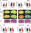
Impaired ability of HO-2-null mice to maintain ATP levels on exposure to 10% O2 for 1 min. (A) Alterations in AMP (AMPwhole), ADP (ADPwhole), ATP (ATPwhole), and energy charge (ECwhole) in the whole brain. The concentrations of adenylates were determined by CE-MS. *P < 0.05 compared with WT normoxia; P < 0.05 compared with HO-2-null normoxia. (B) Representative IMS showing spatial distribution of apparent ATP concentration (ATPapp) and energy charge (ECreg). Note the basal increase in ATP in HO-2-null mice. (Bottom) H&E staining after IMS. cx, cortex; hp, hippocampus. (C) Quantitative analysis of regional ATP concentration and energy charge in WT and HO-2-null mice. *P < 0.05 compared with WT normoxia; P < 0.05 compared with HO-2-null normoxia.

References

    1. Iadecola C, Nedergaard M. Glial regulation of the cerebral microvasculature. Nat Neurosci. 2007;10:1369–1376. - PubMed
    1. Attwell D, et al. Glial and neuronal control of brain blood flow. Nature. 2010;468:232–243. - PMC - PubMed
    1. Ances BM. Coupling of changes in cerebral blood flow with neural activity: What must initially dip must come back up. J Cereb Blood Flow Metab. 2004;24:1–6. - PubMed
    1. Busija DW, Bari F, Domoki F, Louis T. Mechanisms involved in the cerebrovascular dilator effects of N-methyl-d-aspartate in cerebral cortex. Brain Res Brain Res Rev. 2007;56:89–100. - PMC - PubMed
    1. Hoffmeyer HW, Enager P, Thomsen KJ, Lauritzen MJ. Nonlinear neurovascular coupling in rat sensory cortex by activation of transcallosal fibers. J Cereb Blood Flow Metab. 2007;27:575–587. - PubMed

Publication types

MeSH terms

LinkOut - more resources