. 2012 Jan 24;109(4):1329-34.
doi: 10.1073/pnas.1120429109. Epub 2012 Jan 9.

Metabolic click-labeling with a fucose analog reveals pectin delivery, architecture, and dynamics in Arabidopsis cell walls

Affiliations

Metabolic click-labeling with a fucose analog reveals pectin delivery, architecture, and dynamics in Arabidopsis cell walls

Charles T Anderson et al. Proc Natl Acad Sci U S A. .

Abstract

Polysaccharide-rich cell walls are a defining feature of plants that influence cell division and growth, but many details of cell-wall organization and dynamics are unknown because of a lack of suitable chemical probes. Metabolic labeling using sugar analogs compatible with click chemistry has the potential to provide new insights into cell-wall structure and dynamics. Using this approach, we found that an alkynylated fucose analog (FucAl) is metabolically incorporated into the cell walls of Arabidopsis thaliana roots and that a significant fraction of the incorporated FucAl is present in pectic rhamnogalacturonan-I (RG-I). Time-course experiments revealed that FucAl-containing RG-I first localizes in cell walls as uniformly distributed punctae that likely mark the sites of vesicle-mediated delivery of new polysaccharides to growing cell walls. In addition, we found that the pattern of incorporated FucAl differs markedly along the developmental gradient of the root. Using pulse-chase experiments, we also discovered that the pectin network is reoriented in elongating root epidermal cells. These results reveal previously undescribed details of polysaccharide delivery, organization, and dynamics in cell walls.


Conflict of interest statement

The authors declare no conflict of interest.

Figures

Fig. 1.
Fig. 1.
FucAl incorporation in Arabidopsis seedlings. (A–D) Both 0.1 μM Alexa 488-azide (A) and 0.1 μM Alexa 594-azide (C) label seedlings treated with 2.5 μM FucAl for 4 h, but not seedlings treated with 0.01% DMSO for 4 h (B and D). (E and F) FucAl incorporation, but not labeling, is dependent on viability. Seedlings were treated with 2.5 μM FucAl for 4 h before (E) or after (F) fixation in 4% paraformaldehyde for 30 min, followed by labeling with 0.1 μM Alexa 488-azide. Images were recorded with a 10× objective on an epifluorescence microscope using fluorescence filter sets (SI Materials and Methods) or brightfield. Images were collected using identical exposure settings and were not contrast-enhanced. (Scale bar, 100 μm.)
Fig. 2.
Fig. 2.
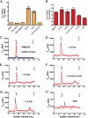
Extraction and characterization of FucAl-labeled cell wall components. (A) Fluorescently labeled FucAl is enriched in CDTA and urea extracts of cell walls. Seedlings were treated with 25 μM FucAl or 0.1% DMSO for 24 h and labeled with Alexa 594-azide. The labeled seedlings were homogenized and cellular components were sequentially extracted. Fluorescence at 585 nm (F585) of 200-μL aliquots of each extract was measured, and the ratio of FucAl-treated to DMSO-treated F585 was calculated. (B) Cell walls prepared from labeled FucAl-treated seedlings were digested with 2.5 U of PME/PG, 1,5-Ara, 1,4-Gal, 1,5-Ara/1,4-Gal, 1,3-Gal, and XEG. Fluorescence was measured as in A and standardized to no enzyme controls (Materials and Methods). Error bars represent SEM from three independent experiments. (C–H) Solubilized material from the indicated digests (red) or no enzyme control (blue) was fractionated on a Sephadex G-75 column, and F585 of 1 mL fractions was measured. Chromatograms are representative of three repetitions of each experiment. Note different scales on y axes of graphs. v0 = void volume, vi = included volume.
Fig. 3.
Fig. 3.
Time-course of FucAl incorporation in elongating root cells. (A–E) Four-day-old seedlings were treated with 2.5 μM FucAl for the indicated times, labeled with Alexa 488-azide, and z series of elongation-zone root epidermal cells were recorded using a spinning disk confocal microscope with a 1.4 NA 100× oil-immersion objective. Images are contrast enhanced maximum projections of the z series. (F) Control seedlings treated with DMSO for 12 h and labeled with Alexa 488-azide show background fluorescence. (G) Maximum projections of z series of elongation-zone root epidermal cells from a 4-d-old seedling treated with 2.5 μM FucAl for 2 h, labeled with Alexa 488-azide, and stained with 0.01% S4B for 30 min. In the merged image on the right, labeled FucAl is green and S4B is red. Lower image shows an x-z projection through the dotted line in a merged z projection and shows that FucAl labeling lies below S4B labeling. (Scale bars, 10 μm.)
Fig. 4.
Fig. 4.
Developmentally distinct patterns of FucAl incorporation in Arabidopsis roots. Four-day-old light-grown Arabidopsis seedlings were treated with 2.5 μM FucAl for 4 h, labeled with 0.1 μM Alexa-488 azide, and imaged by confocal microscopy. (A) Mosaic of maximum projections of a contiguous z series collected starting at the root tip and progressing into the late differentiation zone. (Scale bar, 50 μm.) (B–D) Representative images collected in the late differentiation zone (B), the early differentiation zone (C), and elongation zone (D). (Scale bars 10 μm.) Arrowhead in (B) indicates bright fluorescence associated with a root-hair primordium.
Fig. 5.
Fig. 5.
Pulse-chase analysis of FucAl incorporation. (A) Pulse-labeling protocol. Four-day-old light-grown Col-0 seedlings were treated with 2.5 μM FucAl for 1 h, washed, plated on MS plates lacking FucAl for increasing time periods, and labeled with Alexa 488-azide. (B–F) Maximum projections of z series of root elongation-zone epidermal cells treated with a 1-h pulse of FucAl and chased for the indicated times. Some cells displayed bright fluorescence at the rootward edge after a 12- or 24-h chase (arrowheads in E and F). (Scale bar, 10 μm.) (G) Maximum projection of z series of root differentiation-zone epidermal cells treated with a 1-h pulse of FucAl and chased for 24 h.

References

    1. Somerville C, et al. Toward a systems approach to understanding plant cell walls. Science. 2004;306:2206–2211. - PubMed
    1. Cosgrove DJ. Growth of the plant cell wall. Nat Rev Mol Cell Biol. 2005;6:850–861. - PubMed
    1. Somerville C. Cellulose synthesis in higher plants. Annu Rev Cell Dev Biol. 2006;22:53–78. - PubMed
    1. Reiter WD, Chapple C, Somerville CR. Mutants of Arabidopsis thaliana with altered cell wall polysaccharide composition. Plant J. 1997;12:335–345. - PubMed
    1. Brown DM, et al. Comparison of five xylan synthesis mutants reveals new insight into the mechanisms of xylan synthesis. Plant J. 2007;52:1154–1168. - PubMed

Publication types