

Gut inflammation can boost horizontal gene transfer between pathogenic and commensal Enterobacteriaceae
- PMID: 22232693
- PMCID: PMC3268327
- DOI: 10.1073/pnas.1113246109
Gut inflammation can boost horizontal gene transfer between pathogenic and commensal Enterobacteriaceae
Abstract
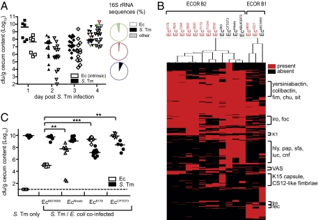
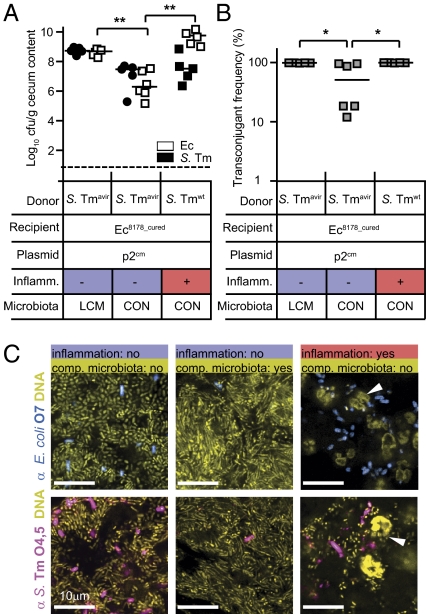
The mammalian gut harbors a dense microbial community interacting in multiple ways, including horizontal gene transfer (HGT). Pangenome analyses established particularly high levels of genetic flux between Gram-negative Enterobacteriaceae. However, the mechanisms fostering intraenterobacterial HGT are incompletely understood. Using a mouse colitis model, we found that Salmonella-inflicted enteropathy elicits parallel blooms of the pathogen and of resident commensal Escherichia coli. These blooms boosted conjugative HGT of the colicin-plasmid p2 from Salmonella enterica serovar Typhimurium to E. coli. Transconjugation efficiencies of ~100% in vivo were attributable to high intrinsic p2-transfer rates. Plasmid-encoded fitness benefits contributed little. Under normal conditions, HGT was blocked by the commensal microbiota inhibiting contact-dependent conjugation between Enterobacteriaceae. Our data show that pathogen-driven inflammatory responses in the gut can generate transient enterobacterial blooms in which conjugative transfer occurs at unprecedented rates. These blooms may favor reassortment of plasmid-encoded genes between pathogens and commensals fostering the spread of fitness-, virulence-, and antibiotic-resistance determinants.
Conflict of interest statement
The authors declare no conflict of interest.
Figures


References
-
- Hehemann JH, et al. Transfer of carbohydrate-active enzymes from marine bacteria to Japanese gut microbiota. (Translated from eng) Nature. 2009;464:908–912. - PubMed
Publication types
MeSH terms
Substances
Associated data
- Actions
- Actions
- Actions
- Actions
- Actions
- Actions
- Actions
- Actions
- Actions
- Actions
- Actions
- Actions
- Actions
- Actions
- Actions
- Actions
- Actions
- Actions
- Actions
- Actions
- Actions
- Actions
- Actions
- Actions
- Actions
- Actions
- Actions
- Actions
- Actions
- Actions
- Actions
- Actions
- Actions
- Actions
- Actions
- Actions
- Actions
- Actions
- Actions
- Actions
- Actions
- Actions
- Actions
- Actions
- Actions
- Actions
- Actions
- Actions
- Actions
- Actions
- Actions
- Actions
- Actions
- Actions
- Actions
- Actions
- Actions
- Actions
- Actions
- Actions
- Actions
- Actions
- Actions
- Actions
- Actions
- Actions
- Actions
- Actions
- Actions
- Actions
- Actions
- Actions
- Actions
- Actions
- Actions
- Actions
- Actions
- Actions
- Actions
- Actions
- Actions
- Actions
- Actions
- Actions
- Actions
- Actions
- Actions
- Actions
- Actions
- Actions
- Actions
- Actions
- Actions
- Actions
- Actions
- Actions
- Actions
- Actions
- Actions
- Actions
- Actions
- Actions
- Actions
- Actions
- Actions
- Actions
- Actions
- Actions
- Actions
- Actions
- Actions
- Actions
- Actions
- Actions
- Actions
- Actions
- Actions
- Actions
- Actions
- Actions
- Actions
- Actions
- Actions
- Actions
- Actions
- Actions
- Actions
- Actions
- Actions
- Actions
- Actions
- Actions
- Actions
- Actions
- Actions
- Actions
- Actions
- Actions
- Actions
- Actions
- Actions
- Actions
- Actions
- Actions
- Actions
- Actions
- Actions
- Actions
- Actions
- Actions
- Actions
- Actions
- Actions
- Actions
- Actions
- Actions
- Actions
- Actions
- Actions
- Actions
- Actions
- Actions
- Actions
- Actions
- Actions
- Actions
- Actions
- Actions
- Actions
- Actions
- Actions
- Actions
- Actions
- Actions
- Actions
- Actions
- Actions
- Actions
- Actions
- Actions
- Actions
- Actions
- Actions
- Actions
- Actions
- Actions
- Actions
- Actions
- Actions
- Actions
- Actions
- Actions
- Actions
- Actions
- Actions
- Actions
- Actions
- Actions
- Actions
- Actions
- Actions
- Actions
- Actions
- Actions
- Actions
- Actions
- Actions
- Actions
- Actions
- Actions
- Actions
- Actions
- Actions
- Actions
- Actions
- Actions
- Actions
- Actions
- Actions
Grants and funding
LinkOut - more resources
Full Text Sources
Other Literature Sources
Molecular Biology Databases
