Review
. 2012 Jan 9;196(1):7-18.
doi: 10.1083/jcb.201108111.

Axon degeneration: molecular mechanisms of a self-destruction pathway

Affiliations
Review

Axon degeneration: molecular mechanisms of a self-destruction pathway

Jack T Wang et al. J Cell Biol. .

Abstract

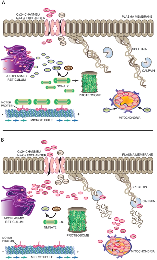
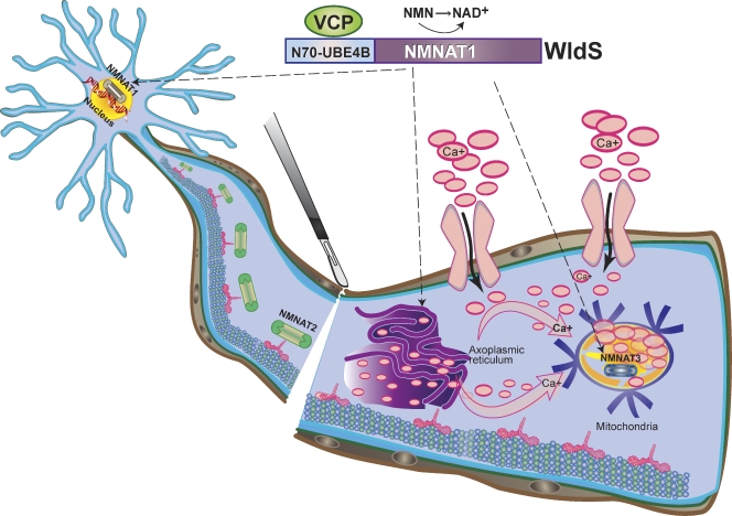
Axon degeneration is a characteristic event in many neurodegenerative conditions including stroke, glaucoma, and motor neuropathies. However, the molecular pathways that regulate this process remain unclear. Axon loss in chronic neurodegenerative diseases share many morphological features with those in acute injuries, and expression of the Wallerian degeneration slow (WldS) transgene delays nerve degeneration in both events, indicating a common mechanism of axonal self-destruction in traumatic injuries and degenerative diseases. A proposed model of axon degeneration is that nerve insults lead to impaired delivery or expression of a local axonal survival factor, which results in increased intra-axonal calcium levels and calcium-dependent cytoskeletal breakdown.


Figures

Figure 1.
Figure 1.
Course of Wallerian axon degeneration. As early as 5–30 min after nerve injury, the axonal segments proximal (left) and distal (right) to the injury site exhibit short-distance acute axon degeneration (AAD), an event that is principally mediated by extracellular Ca2+ influx and activation of the intracellular Ca2+-dependent protease calpain. This event is followed by a slower axonal retraction and formation of axonal bulbs at the injury sites (arrowheads). For the next 24 to 48 h after injury there is a period of relative latency in which the distal axon remains morphologically stable and electrically excitable. Although beading occurs along the distal axon at irregular intervals, there are few signs of physical fragmentation. At more than 72 h after injury, rapid fragmentation and cytoskeletal breakdown occur along the full length of the distal axon, followed by increased glial (consisting primarily of astrocytes, macrophages and, in the PNS, Schwann cells) influx to clear axonal remnants (blue circles) and to possibly promote regenerative attempts by the proximal axon.
Figure 2.
Figure 2.
A molecular model of axon degeneration. (A) In the absence of injury, there is a balance between continuous somal supply via anterograde transport of an axon survival signal such as Nmnat2 and its degradation by the proteasome, resulting in sustained level of the molecule in the axon. Sufficiently high local NAD+ levels, resulting from enzymatic activity of Nmnat2, may keep axonal Ca2+ levels low by regulating the movement of Ca2+ in and out of axonal Ca2+ storage sites such as axoplasmic reticulum and mitochondria through a so-far unidentified mechanism. This regulation of Ca2+ levels in the axoplasm prevents the activation of Ca2+-dependent proteases from cleaving cytoskeletal proteins such as spectrin and preserves the structural and functional integrity of the axon. (B) Nerve injury results in impaired somal supply of Nmnat2 to the axon, resulting in diminished levels of the protein as well as axonal NAD+. Lack of NAD+-dependent regulation of Ca2+ levels lead to increased Ca2+ channel-mediated influx, reverse activity of the Na+/Ca2+ exchanger, or release of Ca2+ from internal storage sites, which together contribute to a catastrophic rise in intra-axonal Ca2+. High Ca2+ levels activate Ca2+-dependent proteases and initiate proteolytic degradation of spectrin and other axonal cytoskeletal components.
Figure 3.
Figure 3.
A molecular model of WldS-mediated axon protection. The WldS fusion protein, consisting of the full-length Nmnat1 and the first 70 amino acids of Ube4b, is predominantly localized in the nucleus; however, it is also expressed in axonal cytoplasm and organelles such as mitochondria (broken arrows denote known neuronal sites of WldS expression) likely due to interaction with the cytoplasmic VCP protein. Expression of either WldS or extranuclear forms of Nmnat1 is sufficient to protect axons from degeneration upon injury, and this may result from substituting for the activity of Nmnat2 protein, which is degraded quickly after nerve injury. The WldS protein may also augment the enzymatic activity of Nmnat3, a mitochondrial Nmnat isoform, to confer axon protection. The combined result of ectopic Nmnat activities in WldS neurons may be less intracellular Ca2+ release from axoplasmic reticulum or greater Ca2+ buffering by the mitochondria via increased NAD+ production in these organelles, leading to overall decrease in intra-axonal Ca2+ levels (pink arrows denote net direction of Ca2+ flux).

References

    1. Adalbert R., Gillingwater T.H., Haley J.E., Bridge K., Beirowski B., Berek L., Wagner D., Grumme D., Thomson D., Celik A., et al. 2005. A rat model of slow Wallerian degeneration (WldS) with improved preservation of neuromuscular synapses. Eur. J. Neurosci. 21:271–277 10.1111/j.1460-9568.2004.03833.x - DOI - PubMed
    1. Alano C.C., Garnier P., Ying W., Higashi Y., Kauppinen T.M., Swanson R.A. 2010. NAD+ depletion is necessary and sufficient for poly(ADP-ribose) polymerase-1-mediated neuronal death. J. Neurosci. 30:2967–2978 10.1523/JNEUROSCI.5552-09.2010 - DOI - PMC - PubMed
    1. Araki T., Sasaki Y., Milbrandt J. 2004. Increased nuclear NAD biosynthesis and SIRT1 activation prevent axonal degeneration. Science. 305:1010–1013 10.1126/science.1098014 - DOI - PubMed
    1. Avery M.A., Sheehan A.E., Kerr K.S., Wang J., Freeman M.R. 2009. Wld S requires Nmnat1 enzymatic activity and N16-VCP interactions to suppress Wallerian degeneration. J. Cell Biol. 184:501–513 10.1083/jcb.200808042 - DOI - PMC - PubMed
    1. Babetto E., Beirowski B., Janeckova L., Brown R., Gilley J., Thomson D., Ribchester R.R., Coleman M.P. 2010. Targeting NMNAT1 to axons and synapses transforms its neuroprotective potency in vivo. J. Neurosci. 30:13291–13304 10.1523/JNEUROSCI.1189-10.2010 - DOI - PMC - PubMed

Publication types

MeSH terms