. 2012 Apr 10;29(6):1197-208.
doi: 10.1089/neu.2011.2261. Epub 2012 Apr 10.

Disruptions in the regulation of extracellular glutamate by neurons and glia in the rat striatum two days after diffuse brain injury

Affiliations

Disruptions in the regulation of extracellular glutamate by neurons and glia in the rat striatum two days after diffuse brain injury

Jason M Hinzman et al. J Neurotrauma. .

Abstract

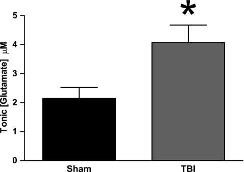
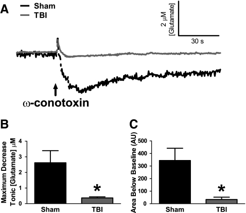
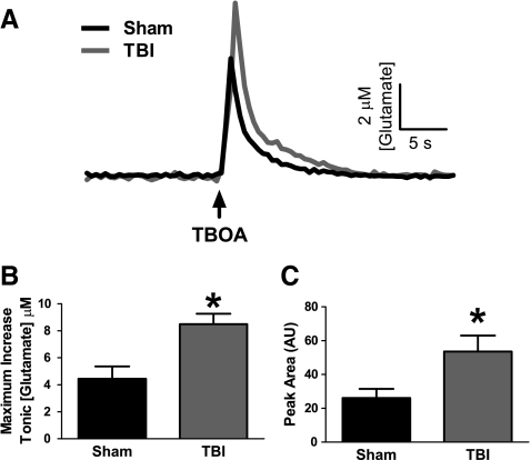
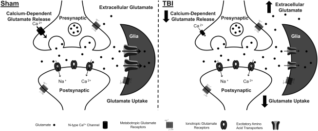
Disrupted regulation of extracellular glutamate in the central nervous system contributes to and can exacerbate the acute pathophysiology of traumatic brain injury (TBI). Previously, we reported increased extracellular glutamate in the striatum of anesthetized rats 2 days after diffuse brain injury. To determine the mechanism(s) responsible for increased extracellular glutamate, we used enzyme-based microelectrode arrays (MEAs) coupled with specific pharmacological agents targeted at in vivo neuronal and glial regulation of extracellular glutamate. After TBI, extracellular glutamate was significantly increased in the striatum by (∼90%) averaging 4.1±0.6 μM compared with sham 2.2±0.4 μM. Calcium-dependent neuronal glutamate release, investigated by local application of an N-type calcium channel blocker, was no longer a significant source of extracellular glutamate after TBI, compared with sham. In brain-injured animals, inhibition of glutamate uptake with local application of an excitatory amino acid transporter inhibitor produced significantly greater increase in glutamate spillover (∼ 65%) from the synapses compared with sham. Furthermore, glutamate clearance measured by locally applying glutamate into the extracellular space revealed significant reductions in glutamate clearance parameters in brain-injured animals compared with sham. Taken together, these data indicate that disruptions in calcium-mediated glutamate release and glial regulation of extracellular glutamate contribute to increased extracellular glutamate in the striatum 2 days after diffuse brain injury. Overall, these data suggest that therapeutic strategies used to regulate glutamate release and uptake may improve excitatory circuit function and, possibly, outcomes following TBI.


Figures

FIG. 1.
FIG. 1.
Increased extracellular glutamate in the striatum of brain-injured rats 2 days after midline fluid percussion brain injury, (n=16, *p<0.05).
FIG. 2.
FIG. 2.
Reduced calcium channel-dependent glutamate release in the striatum of brain-injured rats. (A) Baseline-matched traces of extracellular glutamate demonstrate the average effect of 100 nL local application (↑) of 10 μM ω-conotoxin, N-type calcium channel blocker, on extracellular glutamate in sham (black) and TBI animals (gray). (B) The maximum decrease in extracellular glutamate from local application of ω-conotoxin was significantly smaller after TBI than in sham-injured animals. (C) The extent of reduced extracellular glutamate with respect to time (area under curve) was significantly reduced after TBI compared with sham-injured animals (n=4, *p<0.05).
FIG. 3.
FIG. 3.
Increased glutamate spillover from inhibition of glutamate uptake in the striatum of brain-injured rats. (A) Traces of extracellular glutamate showing the average response from 100 nL local application (↑) of 100 μM dl-threo-β-benzyloxyaspartate (TBOA), excitatory amino acid transporter blocker, in sham (black) and TBI (gray). (B) The maximum increase in extracellular glutamate from local application of TBOA was significantly elevated after TBI compared with sham-injured animals. (C) The extent of higher extracellular glutamate with respect to time (area under curve) was significantly increased after TBI compared with sham-injured animals (n=4, *p<0.05).
FIG. 4.
FIG. 4.
Decreased glutamate clearance in the striatum of brain-injured rats. (A) Representative glutamate signals in the striatum from local application of 500 μM glutamate (↑) in sham (black) and TBI (gray) showed significant decreases in glutamate uptake parameters after TBI. (B) The amplitude of the glutamate signal was similar between sham and TBI groups. (C) Trise, an indicator of glutamate diffusion, was similar between sham and TBI groups. (D) k-1, an indicator of glutamate uptake rate, was significantly reduced after TBI compared with sham-injured animals. (E) T80, an indicator of glutamate clearance, was significantly longer after TBI compared with sham-injured animals (n=8, *p<0.05).
FIG. 5.
FIG. 5.
Schematic depicting the regulation of extracellular glutamate in the rat striatum in sham and brain-injured animals. In sham-injured animals, neurons release glutamate into the extracellular space that is dependent on calcium entry through N-type calcium channels. Once in the extracellular space, glutamate acts on both metabotropic and ionotropic glutamate receptors for signal propagation. Glutamate is removed from the extracellular space by excitatory amino acid transporters, predominantly localized to glia. Glutamate uptake is the primary mechanism for removal of extracellular glutamate and is responsible for regulating the concentration extracellular glutamate. After TBI, we detected post-traumatic disruptions in the regulation of extracellular glutamate by both neurons and glia. In brain-injured animals, calcium-dependent glutamate release via N-type calcium channels was no longer a source of extracellular glutamate. Also, the uptake of glutamate by excitatory amino acid transporters was reduced in brain-injured animals. Post-traumatic decreases in glutamate uptake was probably the primary mechanism responsible for increased extracellular glutamate after TBI.

Similar articles

Cited by

References

    1. Albrecht P. Lewerenz J. Dittmer S. Noack R. Maher P. Methner A. Mechanisms of oxidative glutamate toxicity: the glutamate/cystine antiporter system xc- as a neuroprotective drug target. CNS Neurol. Disord. Drug Targets. 2010;9:373–382. - PubMed
    1. Allen J.W. Ivanova S.A. Fan L. Espey M.G. Basile A.S. Faden A.I. Group II metabotropic glutamate receptor activation attenuates traumatic neuronal injury and improves neurological recovery after traumatic brain injury. J. Pharmacol. Exp. Ther. 1999;290:112–120. - PubMed
    1. Baker D.A. Xi Z.X. Shen H. Swanson C.J. Kalivas P.W. The origin and neuronal function of in vivo nonsynaptic glutamate. J Neurosci. 2002;22:9134–9141. - PMC - PubMed
    1. Battaglia G. Monn J.A. Schoepp D.D. In vivo inhibition of veratridine-evoked release of striatal excitatory amino acids by the group II metabotropic glutamate receptor agonist LY354740 in rats. Neurosci. Lett. 1997;229:161–164. - PubMed
    1. Bond A. Jones N.M. Hicks C.A. Whiffin G.M. Ward M.A. O'Neill M.F. Kingston A.E. Monn J.A. Ornstein P.L. Schoepp D.D. Lodge D. O'Neill M.J. Neuroprotective effects of LY379268, a selective mGlu2/3 receptor agonist: investigations into possible mechanism of action in vivo. J. Pharmacol. Exp. Ther. 2000;294:800–809. - PubMed

Publication types

LinkOut - more resources