. 2012 Jul;18(5):459-68.
doi: 10.1111/j.1601-0825.2011.01893.x. Epub 2012 Jan 11.

Enhanced alveolar bone loss in a model of non-invasive periodontitis in rice rats

Affiliations

Enhanced alveolar bone loss in a model of non-invasive periodontitis in rice rats

J I Aguirre et al. Oral Dis. 2012 Jul.

Abstract

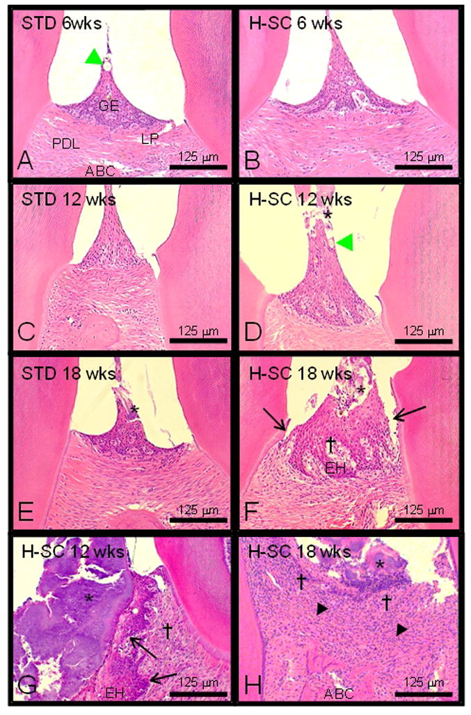
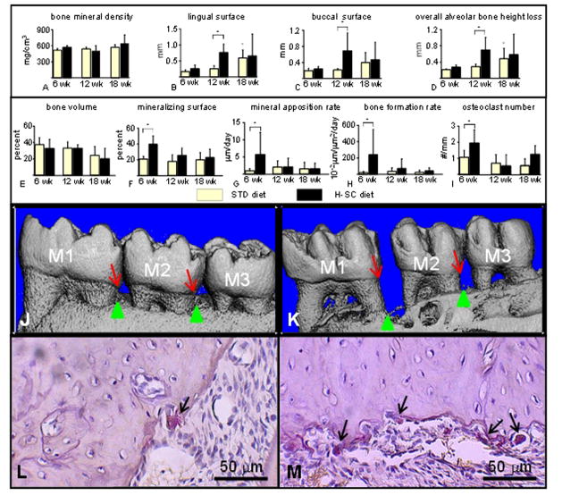
Objective: The rice rat (Oryzomys palustris) develops periodontitis-like lesions when fed a diet rich in sucrose and casein (H-SC). We aimed to establish whether this model can accurately mimic the development of human periodontitis.

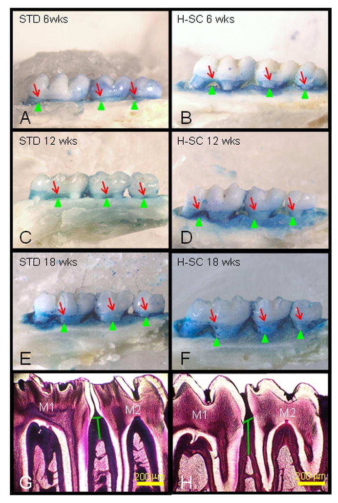
Materials and methods: For this purpose, 28-day-old rice rats (15/group) were assigned to standard (STD) or H-SC diets and sacrificed after 6, 12, and 18 weeks. Jaws were processed for morphometric, histometric, histologic, histomorphometric, and micro-CT analyses.

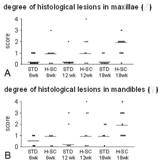
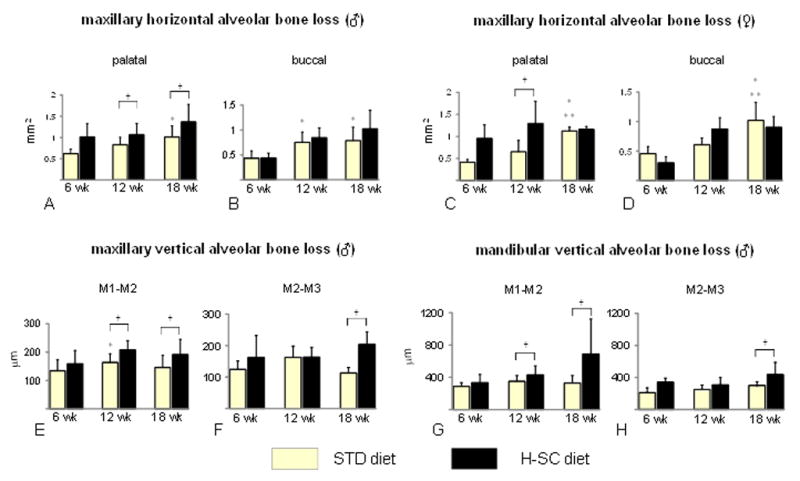
Results: We found a progressive increase in horizontal alveolar bone loss (ABL) with age in maxillae of rats fed the STD diet as determined by morphometry. The H-SC diet exacerbated horizontal ABL at the palatal surface at 12 and 18 weeks. Furthermore, increased vertical ABL was detected in mandibles and maxillae of rats fed the H-SC diet for 12 and/or 18 weeks by histometry and micro-CT. Remarkably, the H-SC diet significantly increased bone remodeling at the interproximal alveolar bone of mandibles from rats fed for 6 weeks, but not in those fed for longer periods.

Conclusions: These findings indicate that the H-SC diet induced a transient increase in alveolar bone remodeling, which is followed by ABL characteristic of moderate periodontitis.


Conflict of interest statement

Conflict of interest

The authors have no conflicts of interest.

Figures

Figure 1
Figure 1
Schematic view of the region of interest (ROI) for the quantitative histomorphometric analysis. Histomorphometric analyses were performed within an area located at the interproximal alveolar bone between the first mandibular molar (M1) and the second mandibular molar (M2). The ROI boundaries are: coronally - a longitudinal line projected through the cementoenamel junction (CEJ) of M1 and M2; apically - a longitudinal line at the level of the apices of the roots of these molars; and mesially and distally - the distal root surface of M1 and the mesial root surface of M2, respectively.
Figure 2
Figure 2
The high sucrose and casein (H-SC) diet induces predominantly slight to moderate periodontal lesions in maxillae and mandibles in rice rats after 12 and/or 18 wks. Degree of periodontal lesions in maxillae of male rats (A) and mandibles of female rats (B) fed the standard (STD) or the H-SC diets for 6, 12 and 18 wks. An inflammation scoring system was used, ranging from 0–4, where 0 corresponds to no histological lesions, 1 to slight, 2 to mild, 3 to moderate and 4 to severe inflammatory lesions affecting some or all of the periodontal structures. Details regarding this scoring system are described in Table 2. Each symbol represents an individual rat. Horizontal lines represent the median for each experimental group.
Figure 3
Figure 3
The high sucrose and casein (H-SC) diet induces increased horizontal and vertical alveolar bone loss (ABL) in rice rats after 12 and/or 18 wks. Maxillary horizontal ABL at the palatal surface of males (A), buccal surface of males (B), palatal surface of females, (C) and buccal surface of females (D). Maxillary vertical ABL at the interproximal space between the first and second molars (M1–M2) in males (E), and between the second and third molars (M2–M3) in males (F) fed the STD and the H-SC diets for 6, 12 and 18 weeks, respectively. Mandibular vertical ABL at the interproximal space of M1–M2 (G) and M2–M3 (H) in male rice rats fed the STD and the H-SC diets for 6, 12 and 18 weeks. An asterisk denotes a significant difference from the STD diet at 6 weeks. Two asterisks denote a significant difference from the STD diet at 12 weeks. A cross (†) above brackets indicates a significant difference from its respective time-matched STD control group. (P < 0.05).
Figure 4
Figure 4. The high sucrose and casein (H-SC) diet increases horizontal and vertical alveolar bone loss (ABL) in rice rats
Comparative images stained with methylene blue to delineate the cementoenamel (CEJ) junction (red arrows) from the maxillary palatal surface of male rice rats fed the standard (STD) or H-SC diets for 6 wks (A and B), 12 wks (C and D) and 18 wks (E and F), respectively. The green arrowheads depict the borders of the alveolar bone crest (ABC). The blue-stained area corresponds to the area of exposed tooth root that can serve as an index of horizontal alveolar bone height. Note the progressive increase in the blue-stained area from rat fed the STD diet for 6 wks (A) to 12 wks (C) and 18 wks (E). Also note that rats fed the H-SC diet exhibited a further increase in the blue surface area, particularly after 12 (D) and 18 wks (F), compared to their time-matched controls (C and E, respectively). Figure H depicts increased mandibular vertical ABL (green vertical line) at the interproximal alveolar bone at molar (M) 1 and M2 in a male rat fed the H-SC diet for 18 wks compared to an age-matched control rat fed the STD diet (G). Methacrylate embedded section stained with en bloc basic fuchsin. Bars = 200 μm (G, H).
Figure 5
Figure 5
The high sucrose and casein (H-SC) diet induces slight to moderate periodontitis in the mandibles of rice rats fed for 12 wks and 18 wks. Comparative photos taken at the interproximal space between the first and second mandibular molars of rice rats fed standard (STD) or H-SC diets for 6 wks (A and B), 12 wks (C and D & G), and 18 wks (E and F & H), respectively. Periodontal lesions were absent or slight in rice rats fed the standard (STD) diet for 6–12 wks and the H-SC diet for 6 wks. A slight degree of inflammation of the periodontium is characterized by slight hyperplasia of the gingival epithelium (GE) with intraepidermal inflammatory cell infiltration and hair shafts or vegetable fibers impacted (green arrowhead) within the gingival epithelium. No substantial changes were observed in the lamina propria (LP), the periodontal ligament (PDL) or the alveolar bone crest (ABC). Periodontal lesions were predominantly slight to moderate (Table 2) in rice rats fed the H-SC diet for 12 wks and 18 wks. Bacterial plaques (*) were frequently observed in rats fed the H-SC diet for 12 and 18 wks, but also in those fed the STD diet for 18 wks (Figures D–H). Figure F illustrates accumulation of bacterial plaque (*), mild migration of the junctional epithelium (black arrow), GE hyperplasia (EH), and mild inflammatory cell infiltration in the LP (†) of a rat fed the H-SC diet for 18 wks. In only 1 of 8 and in 1 of 9 female rats, severe lesions were found in their mandibles after feeding the H-SC diet for 12 and 18 wks, respectively. Figure G illustrates a massive accumulation of bacterial plaque (*) in the interproximal area, disruption of the gingival papilla, EH with migration of the junctional epithelium (black arrow), and inflammatory cell infiltration in the LP (†) of a rat fed the H-SC diet for 12 wks. Figure H shows a severe disruption of the periodontium in one rat fed the H-SC diet for 18 wks. Bacterial plaque (*) is observed in the interproximal area, ulceration of the GE, abundant inflammatory cell infiltration in the LP (†), disruption of the PDL (▲), and alveolar bone osteolysis. Figures were taken at 200X magnification. H&E stain. Bars = 125 μm.
Figure 6
Figure 6
The high sucrose and casein (H-SC) diet increases mandibular vertical alveolar bone loss (ABL) in female rice rats after 12 wks, and increases alveolar bone remodeling at 6 wks but not at 12 and 18 wks in male rice rats. A microCT analysis was performed in the mandibles of female rats fed the standard (STD) and H-SC diets for 6, 12 and 18 weeks (A–D). Bone mineral density (BMD) (A), mandibular vertical ABL at the lingual surface (B), mandibular vertical ABL at the buccal surface (C) and overall alveolar bone height loss (D). All measurements were performed at the interproximal space between the first and second molars (M1–M2). A histomorphometric analysis was performed in the mandibles of male rats fed the standard (STD) and H-SC diets for 6, 12 and 18 weeks (E–I). Bone volume (BV/TV) (E), mineralizing surface (F), mineral apposition rate (G), bone formation rate (H), and osteoclast number (I). All measurements were performed within the region of interest (ROI) described in Figure 1. An asterisk denotes a significant difference from the STD diet at 6 weeks. A cross (†) above brackets indicates a significant difference from its respective time-matched STD control group. (P < 0.05). Comparative reconstructed microCT images taken from mandibles of female rice rats fed the STD (J) or the H-SC diets (K) for 12 weeks, respectively. Note the increased vertical ABL at the lingual surface in the interproximal spaces between molar (M) 1 and M2 and M2–M3 in a rat fed the H-SC diet (K) compared to an age-matched control rat (J). Red arrows depict the cementoenamel junction (CEJ) and green arrows the alveolar bone crest (ABC). Representative photos of histological sections stained for tartrate-resistant acid phosphatase (TRAP) taken at the mandibular interproximal space M1–M2 of a female rice rat fed the STD (L) or H-SC (M) diets for 6 wks, respectively. Note the presence of numerous big multinuclear or mononuclear purple stained osteoclasts (TRAP positive; black arrows) on an scalloped alveolar bone surface in a rat fed the H-SC diet for 6 wks (M) compared to the presence of only one TRAP positive cell (black arrow) on a smoother alveolar bone surface of a rice rat fed the STD diet for 6 wks (L). Figures L and M were taken at 400X magnification. H&E stain. Bars = 50 μm.

References

    1. Allen MR. Animal models of osteonecrosis of the jaw. J Musculoskelet Neuronal Interact. 2007;7 (4):358–360. - PubMed
    1. Allen MR, Burr DB. Three years of alendronate treatment results in similar levels of vertebral microdamage as after one year of treatment. J Bone Miner Res. 2007;22 (11):1759–1765. - PubMed
    1. Armitage GC, Cullinan MP. Comparison of the clinical features of chronic and aggressive periodontitis. Periodontol 2000. 2010;53:12–27. - PubMed
    1. Auskaps A, Gupta O, Shaw J. Periodontal disease in the rice rat. III. Survey of dietary influences. J Nutr. 1957;63 (3):325–343. - PubMed
    1. Bainbridge B, et al. Role of Porphyromonas gingivalis phosphoserine phosphatase enzyme SerB in inflammation, immune response, and induction of alveolar bone resorption in rats. Infect Immun. 2010;78 (11):4560–4569. - PMC - PubMed

Publication types