. 2012 Apr;12(4):867-76.
doi: 10.1111/j.1600-6143.2011.03917.x. Epub 2012 Jan 10.

An obligatory role for lung infiltrating B cells in the immunopathogenesis of obliterative airway disease induced by antibodies to MHC class I molecules

Affiliations

An obligatory role for lung infiltrating B cells in the immunopathogenesis of obliterative airway disease induced by antibodies to MHC class I molecules

N Fukami et al. Am J Transplant. 2012 Apr.

Abstract

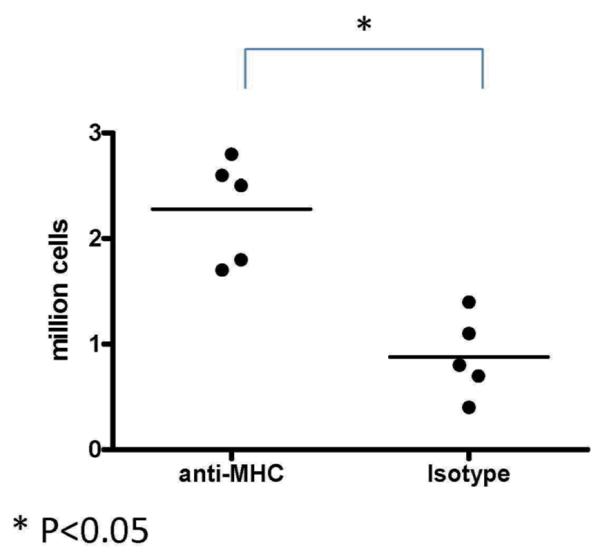
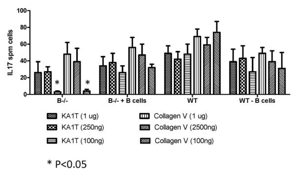
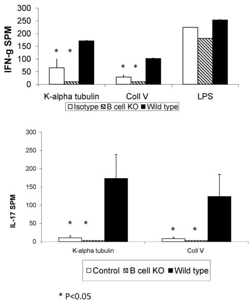
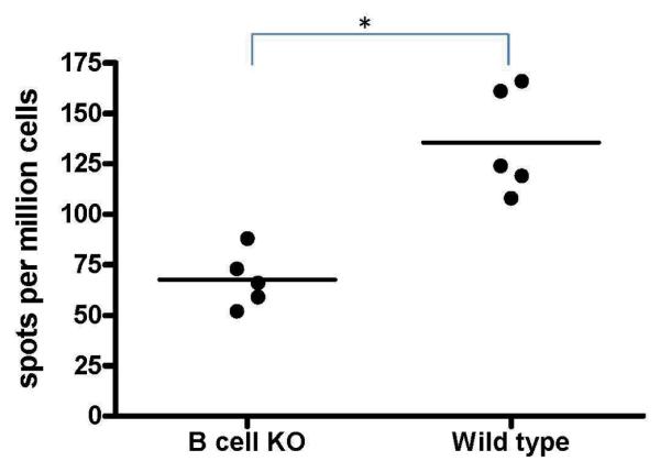
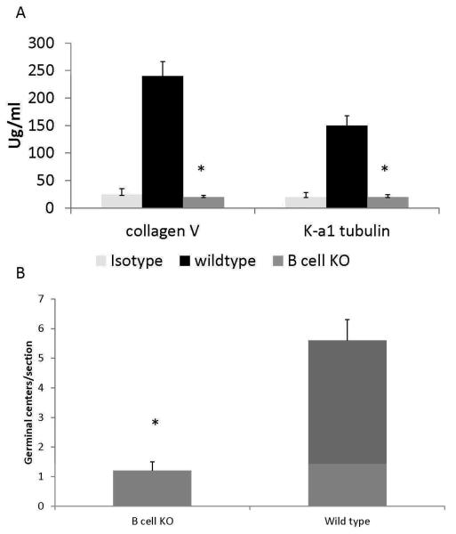
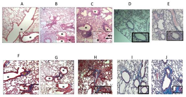
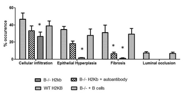
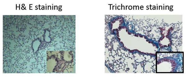
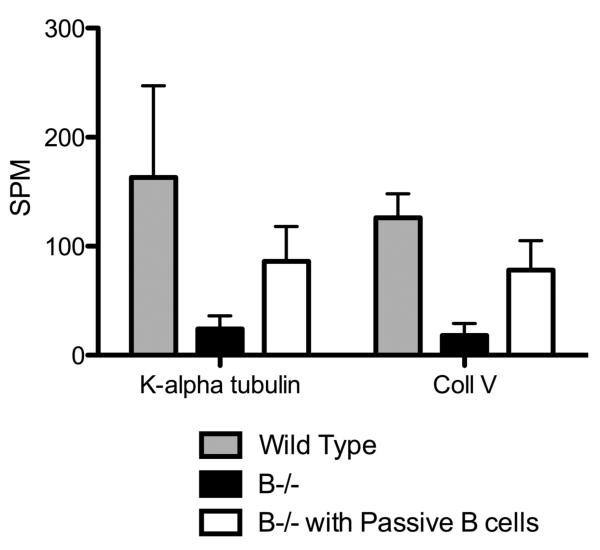
Using a murine model, we demonstrated that endobronchial administration of antibodies (Abs) to major histocompatibility complex (MHC) class I results in cellular infiltration, epithelial metaplasia, fibrosis and obstruction of the small airways (obliterative airway disease [OAD]) mediated predominantly by Th17 responses to self-antigens. This resembles bronchiolitis obliterans syndrome developed following human lung transplantation. Since B cells play a crucial role in induction of autoimmune responses, we defined the role of B cells and its antigen presenting properties in induction of OAD in this study. Anti-MHC class I was administered endobronchially in B(-/-) and wild-type mice. In contrast to wild type, B(-/-) animals did not demonstrate cellular infiltration, epithelial metaplasia and obstruction of airways following anti-MHC. Frequency of K-α1 tubulin and CollagenV-specific IL-17 cells was significantly decreased in B(-/-) mice. As expected, Abs against self-antigens and germinal center formation were not developed in B(-/-) mice. Thus, we conclude that B cells and its antigen presenting capacity play an important role in induction of immune responses to self-antigens and immunopathogenesis of OAD following the administration of anti-MHC. Therefore, strategies to block B-cell and its antigen presenting functions should be considered for preventing the development of chronic rejection.


Figures

Figure 1
Figure 1. Increased infiltration of B cells in lungs of mice administered anti-MHC Abs endobronchially
Anti-H2kb or control (C1.18.4) Ab was administered endobronchially in C57BL/6 mice on days 1, 2, 3. The lungs were harvested on day 4 and analyzed for the number of infiltrating cells by flow cytometry using anti-CD19 Abs. There was a significant increase in the number of B cells in the lung following anti-MHC Ab administration. Represented as mean number of B cells with 5 mice in each group. *Denotes p<0.05
Figure 2
Figure 2. B cell depletion results in decreased IL-17 secreting cells when stimulated with low concentrations of self-antigens
Splenocytes (3×10⁵ per well) from B6.129S2-Igh-6tm1Cgn/J (B−/−) or control C57BL/6 mice (Wild type – WT) were stimulated with 1μg, 250ng and 100 ng of K-α1T or ColV (cells cultured in triplicate for each concentration of antigen) and the frequency of IL-17 secreting cells analyzed using ELISPOT. In another set of experiment B cells from wild type mice were added to the T cells from B6.129S2-Igh-6tm1Cgn/J or B cells were depleted from the splenocytes of wild type mice and stimulated with self-antigens to determine the frequency of IL-17 secreting T cells. The frequency of IL-17 secreting T cells were lower in splenocytes from B6.129S2-Igh-6tm1Cgn/J and wild type mice depleted of B cells (at 100ng antigen concentration) when compared to wild type mice as well as B cells added splenocytes from B6.129S2-Igh-6tm1Cgn/J mice. Frequency of IL-17 secreting cells following stimulation at higher antigen concentration (250ng and 1μg) did not differ between the groups. Results are expressed as mean ± SD spots per million cells observed in 5 mice per group. * Denotes P<0.05
Figure 3
Figure 3. Significant reduction in the pro-inflammatory cytokines and chemokines in B cell deficient mice following anti-MHC Ab administration
B6.129S2-Igh-6tm1Cgn/J (B−/−) or control C57BL/6 mice (Wild type) were treated with anti-MHC class I Abs day 1, 2 and 3. We analyzed the expression levels of cytokines and chemokines in the lungs by quantitative real-time PCR on Day 4. There was a significant reduction in the expression of TNF-α (4.6 folds), IL-6 (6.8 folds), IL-2 (2.7 folds), IFN-γ (8.3 folds) and CXCL12 (14.4 folds) in the lungs of B cell deficient mice when compared to the wild type controls. Data representative of mean ± SD fold change observed, with 5 mice per group.
Figure 4
Figure 4. B cell deficiency results in a significant reduction in the frequency of IFN-γ and IL-17 secreting T cells against K-α1T and ColV in the lung following anti-MHC Ab administration
B6.129S2-Igh-6tm1Cgn/J (B cell KO) or control C57BL/6 mice were treated with anti-MHC class I Abs on day 1, 2, 3, 6 and weekly thereafter. T cells infiltrating the lungs were harvested by collagenase digestion and the frequency of T cells secreting IFN-γ, IL-4 and IL-17 on stimulation with K-α1T and ColV (3×10⁵ cells/well cultured in triplicate for each antigen) were analyzed by ELISPOT. A significant reduction in the frequency of T cells against K-α1T and ColV was observed in B cell deficient mice when compared to controls. IFN-g secreting cells to LPS in vitro did not differ among the groups. Data represented as mean ± SD spots per million cells (spm) of 5 mice per group.
Figure 5
Figure 5. Alloimmune responses are preserved in B cell KO mice albeit of a lower magnitude compared to wild type mice
B6.129S2-Igh-6tm1Cgn/J (B cell KO) or control C57BL/6 (Wild type) mice were immunized twice with 1 million BALB/c splenocytes intraperitoneally at weekly intervals. Frequency of IFN-γ secreting cells that respond to the BALB/c splenocytes were analyzed by ELISPOT. B cell deficient mice immunized with BALB/c splenocytes mounted an IFN-γ response against the mismatched MHC antigens albeit of a lower magnitude when compared to their wild type controls. Represented as mean spots per million cells of 5 mice per group.
Figure 6
Figure 6
A: Decreased auto-Ab production in B cell deficient mice following anti-MHC Ab administration. B6.129S2-Igh-6tm1Cgn/J (B cell KO) or control C57BL/6 (wild type) mice were treated with anti-MHC class I Abs day1, 2, 3, 6 and weekly thereafter. Serum collected at day 30 was analyzed for auto-Abs against K-α1T and ColV by ELISA. Abs to K-α1T and ColV were significantly reduced in B cell deficient mice when compared to wild type following administration of anti-MHC Abs. Represented as mean ± SD μg/mL of 5 mice per group. * Denotes p<0.05 B: Formation of germinal centers is significantly reduced in the B cell deficient mice following administration of anti-MHC Abs when compared to wild type controls. B6.129S2-Igh-6tm1Cgn/J (B cell KO) or control C57BL/6 (wild type) mice were treated with anti-MHC class I Abs day 1, 2, 3, 6 and weekly thereafter. Spleens from the mice were harvested on day 30 and analyzed for the germinal center formation by staining with anti-PNA Abs. Immunohistochemical analysis of the spleen from the wild type mice demonstrated significantly increased number of germinal centers in comparison to B cell deficient mice.
Figure 7
Figure 7. B cell deficiency results in a significant reduction in cellular infiltration around vessel and bronchiole, hyperplasia of the bronchial epithelium and fibrosis
B6.129S2-Igh-6tm1Cgn/J (B cell KO), control C57BL/6 (wild type), and B cell KO mice with passive transfer of 2.5×10⁶ B cells were treated with either anti-MHC class I (anti-H2kb), isotype (C1.184) or nonspecific anti-MHC class I (anti H2kd) Abs on day 1, 2, 3, 6 and weekly thereafter (5 mice in each group). The lungs were harvested on day 30. Formalin preserved tissues were sectioned randomly and analyzed by H&E (a – e) and Masson's trichrome staining (d – i). Images were captured and representative area depicted in the figure. At least 4 out of 5 mice from each group demonstrated similar lesions in various sections of the lung. None of the B cell KO mice treated with anti-H2kb (b, g) demonstrated any significant fibrosis or airway obliteration, with minimal cellular infiltration and epithelial hyperplasia compared to wild type mice treated with anti-H2kd (c, h). B cell KO mice with passive transfer of B cells (e, j) demonstrated lesions similar to wild type mice. Wild type mice treated with non-specific anti-MHC class I (anti H2kd) (d, i) demonstrated no lesions. a,f: B cell KO with isotype control Abs; b,g: B cell KO with anti H2kb; c,h: wild type with anti-H2kb; d,i: wild type with anti-H2kd (non specific anti-MHC); e,j: B cell KO with passive transfer of B cells. B: bronchiole and V: vessels. Blue staining in trichrome stains denotes collagen deposition in the lungs.
Figure 8
Figure 8. Morphometric analysis showing reduced cellular infiltration, epithelial hyperplasia, fibrosis and luminal occlusion in B cell KO mice and in B cell KO mice with passive transfer of auto-Abs
A: B6.129S2-Igh-6tm1Cgn/J (B cell KO), control C57BL/6 (wild type), B cell KO mice with passive transfer of 2.5×10⁶ B cells (purified from splenocytes using B cell enrichment kit, Miltenyi Biotech) and B cell KO mice with passive transfer of 250ng each of Abs to ColV and K-α1T (on day 1) were treated with anti-MHC class I (anti-H2kb Abs on day 1, 2, 3, 6 and weekly thereafter (5 mice in each group). The lungs were harvested on day 30. Formalin preserved tissues were sectioned randomly and analyzed by H&E and Masson's trichrome staining. Images were captured and sections morphometric analysis performed using Optimas software (Media cybernetics). B-cell KO mice demonstrated significantly lesser bronchioles and vessels with cellular infiltration and epithelial hyperplasia. On passively transferring auto-Abs, only some bronchioles and vessels had epithelial hyperplasia or cellular infiltration with absence of any fibrosis when compared to wild type mice or B cell KO mice with passively transferred B cells. % occurrence refers to the percentage of bronchioles/vessels with cellular infiltration, epithelial hyperplasia, fibrosis or luminal occlusion per total number of bronchioles/vessels visualized in the lung sections. Data is mean ± SD of that observed in 5 mice per group. B: H&E and trichrome staining of the lungs. C: ELISPOT analysis of the lung infiltrating T cells specific for K-α1T and ColV in wild type mice, B−/− mice and B−/− mice passively transferred B cells from wild type mice.

Similar articles

Cited by

References

    1. Lange W. Uber eine eigenthumbiche erkankung der kleinen broncheim and bronchiolen. Deutsche Arch Klin Med. 1901;70:342–364.
    1. Jain R, Hachem RR, Morrell MR, Trulock EP, Chakinala MM, Yusen RD, et al. Azithromycin is associated with increased survival in lung transplant recipients with bronchiolitis obliterans syndrome. J Heart Lung Transplant - PMC - PubMed
    1. Sundaresan RS, Trulock EP, Mohanakumar T, Cooper JD, Patterson GA. Prevalence and outcome of bronchiolitis obliterans syndrome after lung transplantation. Washington University Lung Transplant Group. Annals of Thoracic Surgery. 1995;60:1341–1347. - PubMed
    1. Valentine VG, Robbins RC, Berry GJ, Patel HR, Reichenspurner H, Reitz BA, et al. Actuarial survival of heart-lung and bilateral sequential lung transplant recipients with obliterative bronchiolitis. J Heart Lung Transplant. 1996;15:371–383. - PubMed
    1. Smith MA, Sundaresan S, Mohanakumar T, Trulock EP, Lynch JP, Phelan DL, et al. Effect of development of antibodies to HLA and cytomegalovirus mismatch on lung transplantation survival and development of bronchiolitis obliterans syndrome. J Thorac Cardiovasc Surg. 1998;116(5):812–820. - PubMed

Publication types