. 2012 Jun;122(11):535-43.
doi: 10.1042/CS20110523.

Toll-like receptor 4 contributes to blood pressure regulation and vascular contraction in spontaneously hypertensive rats

Affiliations

Toll-like receptor 4 contributes to blood pressure regulation and vascular contraction in spontaneously hypertensive rats

Gisele F Bomfim et al. Clin Sci (Lond). 2012 Jun.

Abstract

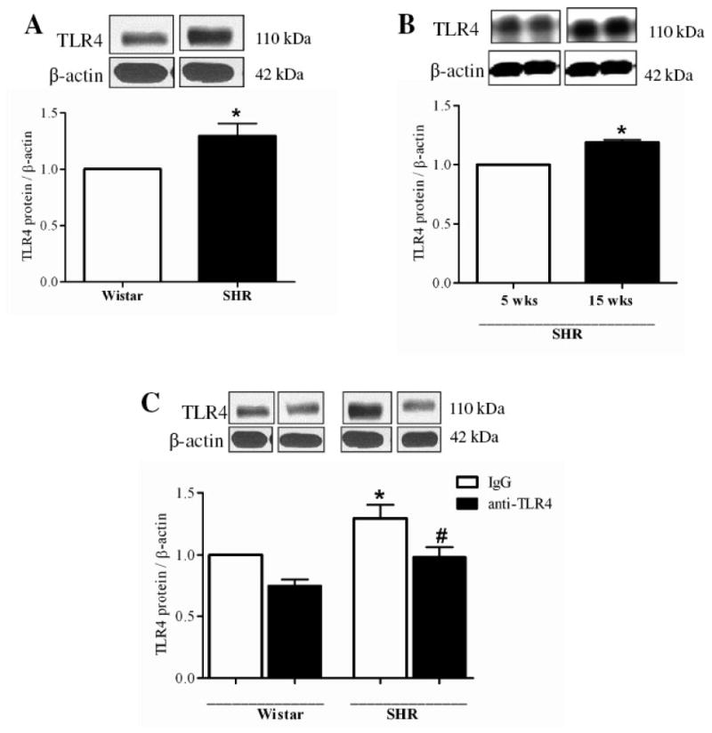
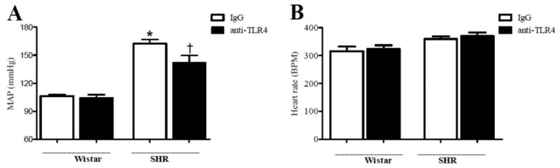
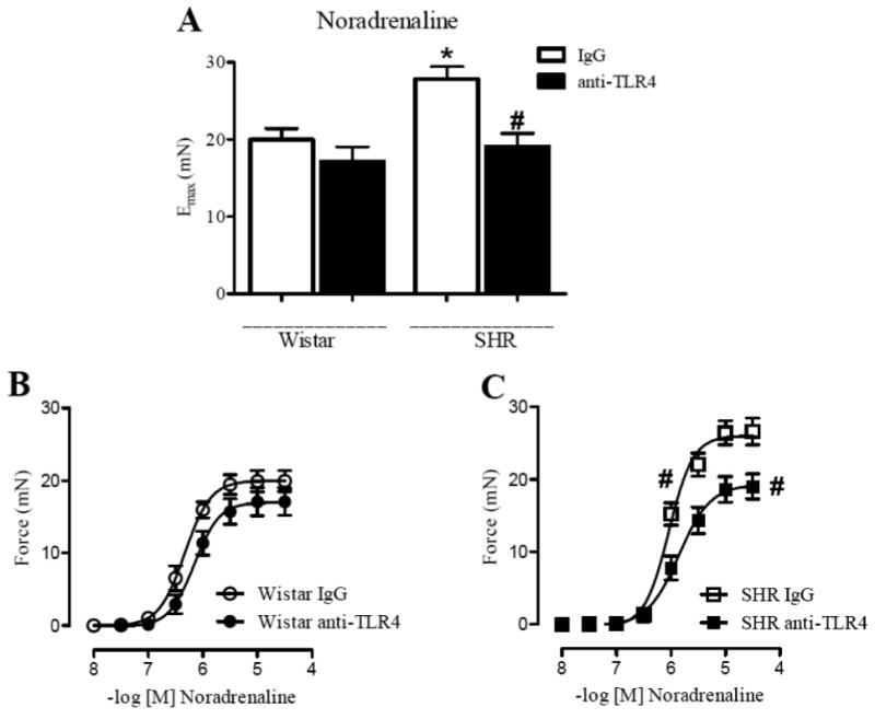
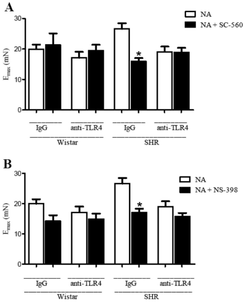
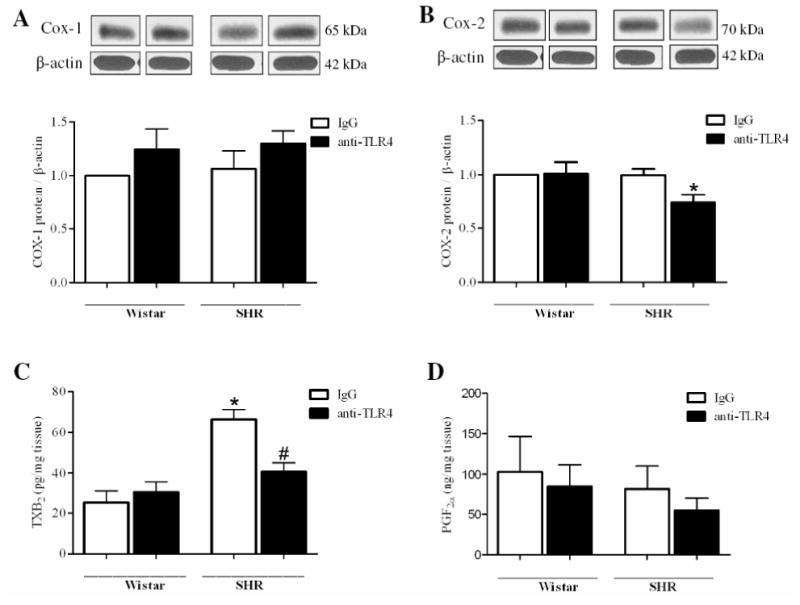
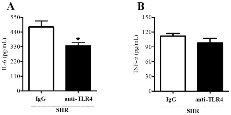
Activation of TLRs (Toll-like receptors) induces gene expression of proteins involved in the immune system response. TLR4 has been implicated in the development and progression of CVDs (cardio-vascular diseases). Innate and adaptive immunity contribute to hypertension-associated end-organ damage, although the mechanism by which this occurs remains unclear. In the present study, we hypothesize that inhibition of TLR4 decreases BP (blood pressure) and improves vascular contractility in resistance arteries from SHR (spontaneously hypertensive rats). TLR4 protein expression in mesenteric resistance arteries was higher in 15-week-old SHR than in age-matched Wistar controls or in 5-week-old SHR. To decrease the activation of TLR4, 15-week-old SHR and Wistar rats were treated with anti-TLR4 (anti-TLR4 antibody) or non-specific IgG control antibody for 15 days (1 μg per day, intraperitoneal). Treatment with anti-TLR4 decreased MAP (mean arterial pressure) as well as TLR4 protein expression in mesenteric resistance arteries and IL-6 (interleukin 6) serum levels from SHR when compared with SHR treated with IgG. No changes in these parameters were found in treated Wistar control rats. Mesenteric resistance arteries from anti-TLR4-treated SHR exhibited decreased maximal contractile response to NA (noradrenaline) compared with IgG-treated SHR. Inhibition of COX (cyclo-oxygenase)-1 and COX-2, enzymes related to inflammatory pathways, decreased NA responses only in mesenteric resistance arteries of SHR treated with IgG. COX-2 expression and TXA2 (thromboxane A2) release were decreased in SHR treated with anti-TLR4 compared with IgG-treated SHR. Our results suggest that TLR4 activation contributes to increased BP, low-grade inflammation and plays a role in the augmented vascular contractility displayed by SHR.


Figures

Figure 1
Figure 1. TLR4 protein expression is augmented in mesenteric resistance arteries from 15 weeks-old SHR compared to Wistar with the same age or to SHR 5-weeks-old. Anti-TLR4 treatment decreased TLR4 protein expression in SHR
(A) TLR4 protein expression was analyzed in mesenteric resistance arteries from 15 week-old Wistar (white bar) and SHR (black bar, and (B) in 5 weeks-old (white bar) and 15 weeks-old (black bar) SHR. (C) We also evaluated TLR4 protein expression in Wistar and SHR rats treated with IgG or anti-TLR4. On top of A, B and C, representative western blot images of TLR4 and P–actin protein expression. Bar graphs show the relative expression of TLR4 after normalization to β-actin expression. Values are means ± SEM, n= 6. *P < 0.05 compared with Wistar (A), 5 wk-old SHR (B) and Wistar IgG (C); # P < 0.05 compared with SHR IgG (C). Statistical test: Student's t test (A and B) and one-way ANOVA (C).
Figure 2
Figure 2. Anti-TLR4 treatment decreased SHR blood pressure
(A) Mean arterial pressure (MAP, mmHg) and (B) heart rate (BPM, beats per minute) were evaluated in Wistar and SHR treated with IgG (white bars) or anti-TLR4 (black bars). Data are expressed as mean ± SEM, n= 6. *P < 0.05 compared with Wistar IgG and #P < 0.05 compared with SHR IgG using a one-way ANOVA.
Figure 3
Figure 3. Noradrenaline-induced vasoconstriction in mesenteric resistance arteries from SHR is attenuated by anti-TLR4 treatment
Maximal response to (A) noradrenaline in mesenteric resistance arteries from Wistar and SHR treated with IgG (white bars) or anti-TLR4 (black bars). Cumulative concentration-response curves to noradrenaline in endothelium-intact mesenteric resistance arteries from (B) Wistar treated with IgG (open circle) or anti-TLR4 (closed circle), and (C) SHR treated with IgG (open square) or anti-TLR4 (closed square). Each point represents the mean ± SEM of maximal response to each concentration, n = 10-12. *P < 0.05 compared with Wistar IgG and # P < 0.05 compared with SHR IgG. Statistical test: one-way ANOVA.
Figure 4
Figure 4. Cox-1 and Cox-2 inhibition decreased the maximal response to noradrenaline only in IgG-treated-SHR mesenteric arteries
Emax to noradrenaline in the absence (white bars) or presence of (A) SC-560 (Cox-1 inhibitor) or (B) NS-398 (Cox-2 inhibitor) (black bars) in mesenteric resistance arteries from Wistar and SHR treated with IgG or anti-TLR4. *P< 0.05 vs. SHR IgG NA. Statistical test: one-way ANOVA.
Figure 5
Figure 5. Anti-TLR4 treatment decreases Cox-2 protein expression and TXB2 release in mesenteric arteries
(A) COX-1 and (B) COX-2 proteins expression in mesenteric resistance arteries from IgG-(white bars) or anti-TLR4-treated (black bars) Wistar and SHR rats. On top, representative western blot images of Cox-1 (A) and Cox-2 (B) protein expression. Bar graphs show the relative expression of Cox-1 and Cox-2 after normalization to β-actin expression. Release of (C) thromboxane B2 and (D) 6-keto-prostaglandin F by mesenteric arteries stimulated with noradrenaline 100 μM. Each bar represents the mean ± SEM, n = 5-6. *P< 0.05. Statistical test: one-way ANOVA.
Figure 6
Figure 6. Anti-TLR4 treatment decreases IL-6 secretion in SHR
Serum levels of (A) IL-6 and (B) TNF-α in IgG- (white bars) and anti-TLR4-treated SHR (black bars). Values are means ± SEM, n = 9. *P < 0.05 vs. IgG-treated SHR. Statistical test: Student`s t test

References

    1. Medzhitov R, Preston-Hurlburt P, Janeway CA., Jr A human homologue of the Drosophila Toll protein signals activation of adaptive immunity. Nature. 1997;388:394–397. - PubMed
    1. Kawai T, Akira S. TLR signaling. Cell Death Differ. 2006;13:816–825. - PubMed
    1. Frantz S, Ertl G, Bauersachs J. Mechanisms of disease: Toll-like receptors in cardiovascular disease. Nat Clin Pract Cardiovasc Med. 2007;4:444–454. - PubMed
    1. Frantz S, Kobzik L, Kim YD, Fukazawa R, Medzhitov R, Lee RT, Kelly RA. Toll4 (TLR4) expression in cardiac myocytes in normal and failing myocardium. J Clin Invest. 1999;104:271–280. - PMC - PubMed
    1. Pasterkamp G, Van Keulen JK, De Kleijn DP. Role of Toll-like receptor 4 in the initiation and progression of atherosclerotic disease. Eur J Clin Invest. 2004;34:328–334. - PubMed

Publication types

MeSH terms