Review
. 2012 Mar 1;4(3):a009662.
doi: 10.1101/cshperspect.a009662.

Synaptic neurotransmitter-gated receptors

Affiliations
Review

Synaptic neurotransmitter-gated receptors

Trevor G Smart et al. Cold Spring Harb Perspect Biol. .

Abstract

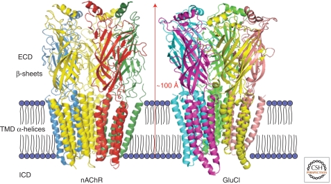
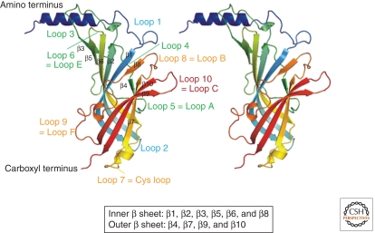
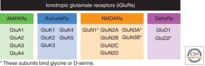
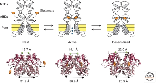
Since the discovery of the major excitatory and inhibitory neurotransmitters and their receptors in the brain, many have deliberated over their likely structures and how these may relate to function. This was initially satisfied by the determination of the first amino acid sequences of the Cys-loop receptors that recognized acetylcholine, serotonin, GABA, and glycine, followed later by similar determinations for the glutamate receptors, comprising non-NMDA and NMDA subtypes. The last decade has seen a rapid advance resulting in the first structures of Cys-loop receptors, related bacterial and molluscan homologs, and glutamate receptors, determined down to atomic resolution. This now provides a basis for determining not just the complete structures of these important receptor classes, but also for understanding how various domains and residues interact during agonist binding, receptor activation, and channel opening, including allosteric modulation. This article reviews our current understanding of these mechanisms for the Cys-loop and glutamate receptor families.


Figures

Figure 1.
Figure 1.
Architecture of Cys-loop receptors. Structure of (left) Torpedo nAChR solved from electron microscopic images at the 4 Å level (pdb 2BG9) (Unwin 2005) and (right) the C. elegans glutamate-gated anion channel at 3.3 Å (pdb 3RIA) (Hibbs and Gouaux 2011). The pentameric subunit assembly and secondary structure are shown for the extracellular domain (ECD) and transmembrane domain (TMD). The ECDs are composed of inner and outer β-sheets with an α-helix, and each subunit’s TMD is formed by four α-helices (M1–M4). Note that for nAChR, the intracellular (MA) helices preceding M4 are omitted, whereas the M3–MA stretch is disordered and is thus not included in the structure. For the GluCl structure, the M3–M4 domain is replaced by a tripeptide, A-G-T (Hibbs and Gouaux 2011).
Figure 2.
Figure 2.
Synaptic view of a Cys-loop receptor. Looking across from the presynaptic terminal over to the postsynaptic membrane, an image of the structure for a typical Cys-loop receptor is shown. This is generated from the atomic resolution structure for AChBP (pdb 2BYQ) (Hansen et al. 2005) for the ECD, linked to the transmembrane domains taken from images of GLIC (Bocquet et al. 2009). (A) The five subunits form a pseudosymmetrical ring with interfacial binding sites between principal (P, +) and complementary (C, −) binding faces. Note the central aqueous pathway for ion conduction. (B) A cut-away slab from A depicts the loop C structures on each subunit and the relative stoichiometry for a muscle nAChR and neuronal GABAAR. The identity of the subunits and neurotransmitter-binding sites are illustrated. (C) Further cut-away to reveal the tops of the TMDs showing M2 lining the ion channel and the support formed by M1, M3, and M4.
Figure 3.
Figure 3.
Structure of the extracellular domain. Side view of two adjacent subunits of the AChBP. The positions of the binding loops and other loops from the amino terminus to the carboxyl terminus that adjoins the pre-M1 domain in Cys-loop receptors are shown. β-strands that form the inner and outer β-sheets are also indicated with labeling according to Brejc et al. (2001).
Figure 4.
Figure 4.
Cys-loop receptor ion channel domain. The M2 domains of several Cys-loop receptors are aligned; those residues that are exposed and lining the channel lumen are boxed. The structural model shows the M2 domains of open GLIC and closed ELIC. For GLIC, the β1-2 loop (green) moves downward and outwardly displaces the M2–M3 linker (blue) hauling the M2 domains (blue) upward and tilting outward to open the channel compared with ELIC (pink and red).
Figure 5.
Figure 5.
The four families of iGluR subunits. Each subunit is encoded by a distinct gene. There is no known mixing of subunits between families.
Figure 6.
Figure 6.
The tetrameric structure of the AMPA GluA2 receptor. (Left) X-ray crystal structure of the AMPA GluA2 homotetrameric receptor (Sobolevsky et al. 2009). Each subunit is in a different color. The tetramer shows a typical layer organization with at the “top” the amino-terminal domains (ATDs or NTDs), at the “bottom” the transmembrane domain (TMD) where the ion channel sits, and sandwiched between the two the agonist-binding domains (ABDs or S1S2 domains) binding glutamate (or glycine/d-serine). (Right) Subunit non-equivalence. α-Carbon traces of subunit A and subunit B with the ABDs similarly oriented. Note the striking difference in overall domain orientation between the two subunits.
Figure 7.
Figure 7.
Structural mechanism of iGluR activation and desensitization. A single dimer is represented; a full receptor is a tetramer made of two such dimers. (Below) The crystal structures of the GluA2 ABD dimer in conformations that correspond to the resting state (no ligand bound; pdb code 1FT0), the active state (glutamate-bound; pdb code 1FTJ), and the desensitized state (pdb code 2I3V). The distances between the two protomers, at the top of the upper lobes (green spheres; dimer interface) and at the bottom of the lower lobes (black spheres; connections to the transmembrane segments), are indicated (Armstrong et al. 2006).
Figure 8.
Figure 8.
Allosteric modulation of iGluRs. (A) Negative allosteric modulation of NMDARs by extracellular zinc. The GluN2A and GluN2B NTDs form subunit-specific inhibitory zinc-binding sites. (Right) Inhibition by nanomolar zinc concentrations of GluN1/GluN2A responses (adapted from Paoletti et al. 2000). (B) Positive allosteric modulation of AMPARs by cyclothiazide (CTZ). CTZ binds and stabilizes the ABD dimer interface. (Right) CTZ blocks desensitization of GluA2 receptors (Sun et al. 2002).

Similar articles

Cited by

References

    1. Absalom NL, Lewis TM, Kaplan W, Pierce KD, Schofield PR 2003. Role of charged residues in coupling ligand binding and channel activation in the extracellular domain of the glycine receptor. J Biol Chem 278: 50151–50157 - PubMed
    1. Alexander SP, Mathie A, Peters JA 2008. Guide to receptors and channels (GRAC), 3rd ed. Br J Pharmacol 153 (Suppl 2): S1–S209 - PMC - PubMed
    1. Amin J, Weiss DS 1996. Insights into the activation mechanism of ρ1 GABA receptors obtained by coexpression of wild type and activation-impaired subunits. Proc R Soc Lond B Biol Sci 263: 273–282 - PubMed
    1. Araud T, Wonnacott S, Bertrand D 2010. Associated proteins: The universal toolbox controlling ligand gated ion channel function. Biochem Pharmacol 80: 160–169 - PubMed
    1. Arevalo E, Chiara DC, Forman SA, Cohen JB, Miller KW 2005. Gating-enhanced accessibility of hydrophobic sites within the transmembrane region of the nicotinic acetylcholine receptor’s δ-subunit. A time-resolved photolabeling study. J Biol Chem 280: 13631–13640 - PubMed

MeSH terms

Substances

LinkOut - more resources