. 2012 Mar 15:205:18-28.
doi: 10.1016/j.neuroscience.2011.12.049. Epub 2012 Jan 3.

Glutamatergic input-output properties of thalamic astrocytes

Affiliations

Glutamatergic input-output properties of thalamic astrocytes

T M Pirttimaki et al. Neuroscience. .

Abstract

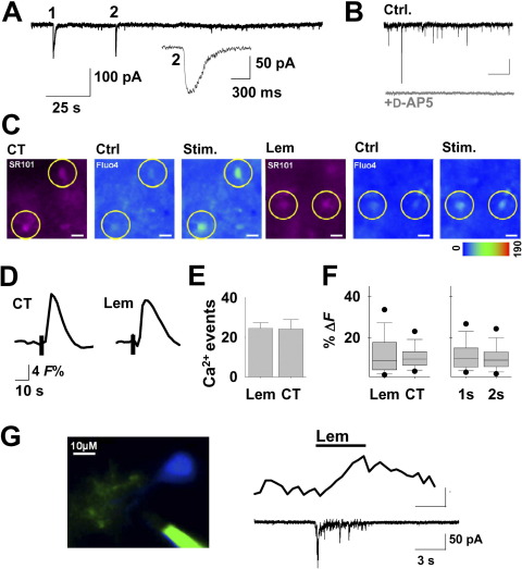
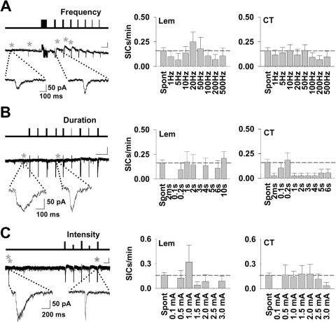
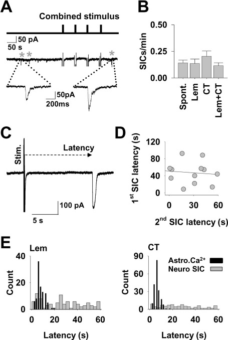
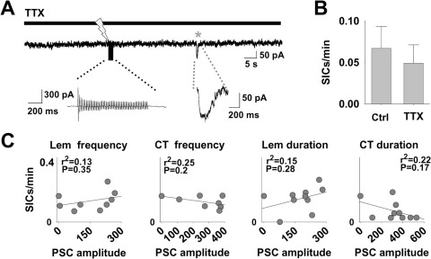
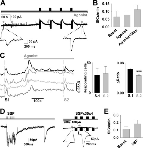
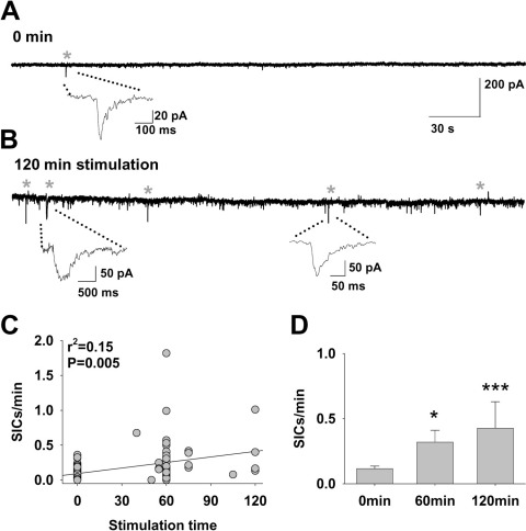
Astrocytes in the somatosensory ventrobasal (VB) thalamus of rats respond to glutamatergic synaptic input with metabotropic glutamate receptor (mGluR) mediated intracellular calcium ([Ca²⁺](i)) elevations. Astrocytes in the VB thalamus also release the gliotransmitter (GT) glutamate in a Ca²⁺-dependent manner. The tripartite synapse hypothesis posits that astrocytic [Ca²⁺](i) elevations resulting from synaptic input releases gliotransmitters that then feedback to modify the synapse. Understanding the dynamics of this process and the conditions under which it occurs are therefore important steps in elucidating the potential roles and impact of GT release in particular brain activities. In this study, we investigated the relationship between VB thalamus afferent synaptic input and astrocytic glutamate release by recording N-methyl-D-aspartate (NMDA) receptor-mediated slow inward currents (SICs) elicited in neighboring neurons. We found that Lemniscal or cortical afferent stimulation, which can elicit astrocytic [Ca²⁺](i) elevations, do not typically result in the generation of SICs in thalamocortical (TC) neurons. Rather, we find that the spontaneous emergence of SICs is largely resistant to acute afferent input. The frequency of SICs, however, is correlated to long-lasting afferent activity. In contrast to short-term stimulus-evoked GT release effects reported in other brain areas, astrocytes in the VB thalamus do not express a straightforward input-output relationship for SIC generation but exhibit integrative characteristics.


Figures

Fig. 1
Fig. 1
Thalamic afferent stimulation activates astrocytes and neurons. (A) Recording from a TC neuron in presence of TTX (1 μM) showing two spontaneous slow inward currents (SICs). SIC 2 is shown expanded. (B) Representative trace showing spontaneous SIC (black, top) sensitivity to d-AP5 (50 μM) (grey, below). (C) First three panels to the left show a slice area loaded with SR101 (magenta images on the left) and Fluo-4 A.M. Circled SR101-positive astrocytes exhibit Ca2+ elevations to CT input (pseudocolor images to the right). Next three panels to the right display a similar experiment to Lemniscal afferent stimulation. (Scale bars: 10 μm). (D) Traces showing averages of 16 CT- and 33 Lemniscal-evoked [Ca2+]i responses to the afferent stimulations. (E) Bar graph showing the mean number of responsive astrocytes within the image field of 444 μm×341 μm (Lem, n=4 slices; CT, n=8). (F) Box plot showing the scatter of relative intensity changes for Lemniscal (n=99 [Ca2+]i responses) and CT (n=194) stimulation, and for 1 (n=149) and 2 s (n=144) train durations. (G) Panel on the left shows confocal image of Alexa 564-filled TC neuron (blue) and astrocyte (green). To the right: recording from the filled cells in showing that 5-s synaptic stimulation of Lemniscal pathway (top bar) elicits [Ca2+]i elevations (middle) in astrocytic processes simultaneously to neuronal EPSC (bottom). For interpretation of the references to color in this figure legend, the reader is referred to the Web version of this article.
Fig. 2
Fig. 2
Synaptic stimulation does not evoke SICs. (A) Line diagram (top) illustrates the protocol of the pattern of episodes of 50 stimuli delivered at different frequencies (1–500 Hz) to CT input. The responses of the TC neuron are shown in the trace below, with SICs indicated by asterisks (Scale bar: 50 s, 50 pA). Expanded example SICs are shown beneath. (Right) Bar graphs show the relationship of mean frequency of SICs (SICs/min) to different stimulus frequencies for the Lemniscal (n=21 neurons) and CT (n=30) inputs. (B) Line diagram (top) illustrates the protocol of the pattern of stimulation delivered at different duration (2 ms–6 s) to CT input. The responses of the TC neuron are shown in the example trace below (Scale bar: 60 s, 50 pA), with SICs indicated by asterisks and examples are expanded beneath. (Right) Bar graphs summarizing the mean frequencies (Lem, n=23; CT, n=40). (C) Line diagram (top) illustrates the protocol of the pattern of stimulation delivered at different intensities (0.5–3 mA) to CT input. The responses of the TC neuron are shown in the example trace below, with SICs indicated by asterisks (Scale bar: 25 s, 50 pA). Expanded example SICs are shown beneath. (Right) Bar graphs show the relationship of mean frequency of SICs to different stimulus intensities (Lem, n=25; CT, n=28). Dashed line in bar graphs indicates mean spontaneous SIC frequency. The neuronal post synaptic currents and stimulation artefacts (vertical lines) are truncated for clarity in (A–C).
Fig. 3
Fig. 3
SIC timing is independent of synaptic stimulation. (A) Line diagram (top) with vertical bars representing simultaneous Lemniscal and CT stimulus episodes. Below is an example trace showing corresponding synaptic responses (vertical lines represent stimulus artefacts and inward currents, the neuronal post synaptic current is truncated for clarity) and SICs (marked with asterisks). (B) Bar graph summarizing the SIC frequency during different stimulus protocols (n=27 neurons). (C) Example of a 0.2-s-long stimulus at 50 Hz delivered to the CT afferent with a SIC following within a few seconds, illustrating the measurement of SIC latency. (D) Scatter plot showing the latency of SICs following a stimulus against the latency of a second SIC to a subsequent stimulus (r2=0.008). (E) Frequency histograms showing the distribution of SIC latencies (grey bars) and Ca2+ delays (black bars) (n=12 slices) following Lemniscal (left) (n=96 neurons) and CT stimulation (right) (n=125 neurons).
Fig. 4
Fig. 4
SIC frequency is not correlated to post-synaptic glutamate effect. (A) Trace of a recording following the addition of TTX which blocks elicited PSC to afferent stimulation (lightning bolt) and only results in a stimulus artefact (expanded below). A spontaneous SIC (grey asterisk) occurs during the recording (expanded below). (B) Mean SIC frequency during control stimulation and following the addition of TTX (n=10 neurons). (C) Mean SIC frequency plotted against the mean PSC amplitude for the different stimulus protocols (CT analysis: n=125 neurons, Lem: n=96 neurons).
Fig. 5
Fig. 5
Physiological modulators or state-dependent stimulation do not increase SIC frequency. (A) Line diagram (top) illustrating the pattern and timing of episodes of 50 stimuli delivered to CT and Lem afferents in the presence of the exogenous agonists carbachol (50 μM) and isoproterenol (50 μM). The responses of the TC neuron are shown in the example trace below, with SICs indicated by asterisks. Expanded example SICs are shown beneath. (B) Bar graph summarizing the SIC frequency for different conditions (Spont, n=12; Agonist, n=16 recordings). (C) Ratio traces from four example astrocytes in an imaged slice showing responses to a 2 s 50 Hz combined afferent input in control conditions (S1) and following exposure to isoproterenol/carbachol (S2) (n=119 astrocytes, 4 slices). Bar graphs to the right illustrate the number of responding astrocytes and magnitude of ratio change to the stimuli in the two conditions. (D) Leftmost: line diagram (top) illustrating the SSP stimulation pattern. Trace below shows the post synaptic current in response to SSP stimulation (left) followed with a delayed SIC (right). To the right: line diagram (top) illustrating the 4 repeated blocks of 30 SSP patterns with recorded current trace below. SICs are asterisked and expanded beneath. (E) Bar graph showing the mean SIC frequency before and during the SSP protocols (n=23 recordings). The neuronal post synaptic current and stimulation artefacts (vertical lines) are truncated for clarity in (C) and (E) (right).
Fig. 6
Fig. 6
Integration of afferent input increases spontaneous SIC frequency. (A) Trace of a recording from an un-stimulated control slice (SICs indicated with asterisks). (B) Trace of a recording from a TC neuron in a slice following a 120 min of prolonged intermittent stimulation. (C) Scatter plot showing a positive correlation of SIC frequency with intermittent stimulus duration. (D) Mean SIC frequency following different intermittent stimulus durations (0 min, n=26; 60 min, n=19; 120 min, n=4 neurons). Statistical significance is presented as * P<0.05, ** P<0.01, or *** P<0.005.

Similar articles

Cited by

References

    1. Agulhon C., Fiacco T.A., McCarthy K.D. Hippocampal short- and long-term plasticity are not modulated by astrocyte Ca2+ signaling. Science. 2010;327:1250–1254. - PubMed
    1. Angulo M.C., Kozlov A.S., Charpak S., Audinat E. Glutamate released from glial cells synchronizes neuronal activity in the hippocampus. J Neurosci. 2004;24:6920–6927. - PMC - PubMed
    1. Araque A., Li N., Doyle R.T., Haydon P.G. SNARE protein-dependent glutamate release from astrocytes. J Neurosci. 2000;20:666–673. - PMC - PubMed
    1. Araque A., Martín E.D., Perea G., Arellano J.I., Buño W. Synaptically released acetylcholine evokes Ca2+ elevations in astrocytes in hippocampal slices. J Neurosci. 2002;22:2443–2450. - PMC - PubMed
    1. Araque A., Parpura V., Sanzgiri R.P., Haydon P.G. Tripartite synapses: glia, the unacknowledged partner. Trends Neurosci. 1999;22:208–215. - PubMed

Publication types