. 2012 Jun;20(6):639-44.
doi: 10.1038/ejhg.2011.250. Epub 2012 Jan 11.

A large duplication involving the IHH locus mimics acrocallosal syndrome

Affiliations

A large duplication involving the IHH locus mimics acrocallosal syndrome

Memnune Yuksel-Apak et al. Eur J Hum Genet. 2012 Jun.

Abstract

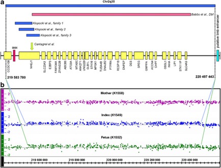
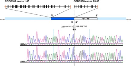
Indian hedgehog (Ihh) signaling is a major determinant of various processes during embryonic development and has a pivotal role in embryonic skeletal development. A specific spatial and temporal expression of Ihh within the developing limb buds is essential for accurate digit outgrowth and correct digit number. Although missense mutations in IHH cause brachydactyly type A1, small tandem duplications involving the IHH locus have recently been described in patients with mild syndactyly and craniosynostosis. In contrast, a ∼600-kb deletion 5' of IHH in the doublefoot mouse mutant (Dbf) leads to severe polydactyly without craniosynostosis, but with craniofacial dysmorphism. We now present a patient resembling acrocallosal syndrome (ACS) with extensive polysyndactyly of the hands and feet, craniofacial abnormalities including macrocephaly, agenesis of the corpus callosum, dysplastic and low-set ears, severe hypertelorism and profound psychomotor delay. Single-nucleotide polymorphism (SNP) array copy number analysis identified a ∼900-kb duplication of the IHH locus, which was confirmed by an independent quantitative method. A fetus from a second pregnancy of the mother by a different spouse showed similar craniofacial and limb malformations and the same duplication of the IHH-locus. We defined the exact breakpoints and showed that the duplications are identical tandem duplications in both sibs. No copy number changes were observed in the healthy mother. To our knowledge, this is the first report of a human phenotype similar to the Dbf mutant and strikingly overlapping with ACS that is caused by a copy number variation involving the IHH locus on chromosome 2q35.


Figures

Figure 1
Figure 1
Phenotypical characteristics of patients K1549 and K1552. (a, b) Craniofacial abnormalities of the index patient as an infant, note the broad forehead, hypertelorism, epicanthic folds, down slanting palpebral fissures, depressed and broad nasal root, short nose with anteverted nostrils, long philtrum and dysplastic, low-set and posterior rotated small ears; (c) patient at 11 years of age; (d) and (e) brain MRI of index patient; (fh) hand and foot malformations of the index patient, note the polydactyly and cutaneous syndactyly; (ik) radiographs of hand and foot malformations of the index patient; (ln) female fetus (K1552) showing similar clinical features as seen in the index patient.
Figure 2
Figure 2
Genomic overview of the duplicated region on chromosome 2q35. (a) Duplications from our study and as published by Klopocki et al are shown in blue, the doublefoot deletion is depicted in pink. The translocation breakpoint in NHEJ1 described by Cantagrel et al is indicated by a green arrow. Genes within the duplicated regions are marked in yellow. The position of IHH is highlighted in red. The putative limb enhancer on chromosome 2q35 is shown in turquoise. Genome positions are given according to hg18 assembly at the UCSC Genome Browser; (b) results of quantitative SNP analysis for patients K1549 and K1552. K1549 and K1552 have a large amplification (CN-State 3) on chromosome 2 with a size of ∼900 kb. The x axis indicates the physical position of the chromosome 2q35. The y axis indicates the log2 ratio of each analyzed SNP in the region. The color reproduction of this figure is available at the European Journal of Human Genetics journal online.
Figure 3
Figure 3
CNV analysis of the IHH locus. Positions 1–4 correspond to the analyzed family (black), patients K1549 and K1552 show values representing three copies of IHH. Positions 5–16 show examples of the control cohort (gray), 17–20 show normalized standard values of a control DNA with different amounts (5 ng, 10 ng, 15 ng and 20 ng, respectively; light blue). Positions 21–24 represent the theoretically expected values of one, two, three and four IHH gene copies (dark blue). The color reproduction of this figure is available at the European Journal of Human Genetics journal online.
Figure 4
Figure 4
Illustration of identified tandem duplications. Schematic overview of genomic position of the analyzed tandem duplication. The duplicated segment is depicted in dark blue, the duplication is shown in dotted blue. Chromatograms of K1549 and K1552 show the identical breakpoints proving tandem orientation of the duplication. The break point is designated by a black line. The genomic positions of the 5′ and 3′ break points are given, and the corresponding bases are indicated by arrows. The 5′ breakpoint is located within exon 25 of CCDC108 (schematic presentation on top) and the intact and the partially duplicated copy of CCDC108 are displayed. The color reproduction of this figure is available at the European Journal of Human Genetics journal online.

References

    1. Zeller R, López-Rios J, Zuniga A. Vertebrate limb development: moving towards integrative analysis of organogenesis. Nat Rev Genet. 2009;10:845–858. - PubMed
    1. Karp SJ, Schipani E, St-Jaques B, Hunzelmann J, Kronenberg H, McMahon AP. Indian hedgehog coordinates endochondral bone growth and morphogenesis via parathyroid hormone related-protein-dependent and –independent pathways. Development. 2000;127:543–548. - PubMed
    1. Long F, Chung UI, Ohba S, McMahon J, Kronenberg HM, McMahon AP. Ihh signaling is directly required for the osteoblast lineage in the enchondral skeleton. Development. 2004;131:1309–1328. - PubMed
    1. Zhou J, Meng J, Guo S, et al. IHH and FGF8 coregulate elongation of digit primordia. Biochem Biophys Res Commun. 2007;363:513–518. - PubMed
    1. Gao B, Guo J, She C, et al. Mutations in IHH, encoding Indian hedgehog, cause brachydactyly Type A-1. Nat Genet. 2001;28:386–388. - PubMed

Publication types