. 2012 Jul;32(7):1310-6.
doi: 10.1038/jcbfm.2011.186. Epub 2012 Jan 11.

The two pathophysiologies of focal brain ischemia: implications for translational stroke research

Affiliations

The two pathophysiologies of focal brain ischemia: implications for translational stroke research

Konstantin-Alexander Hossmann. J Cereb Blood Flow Metab. 2012 Jul.

Abstract

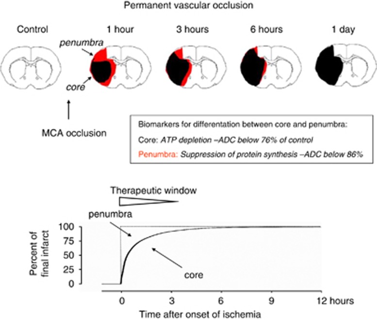
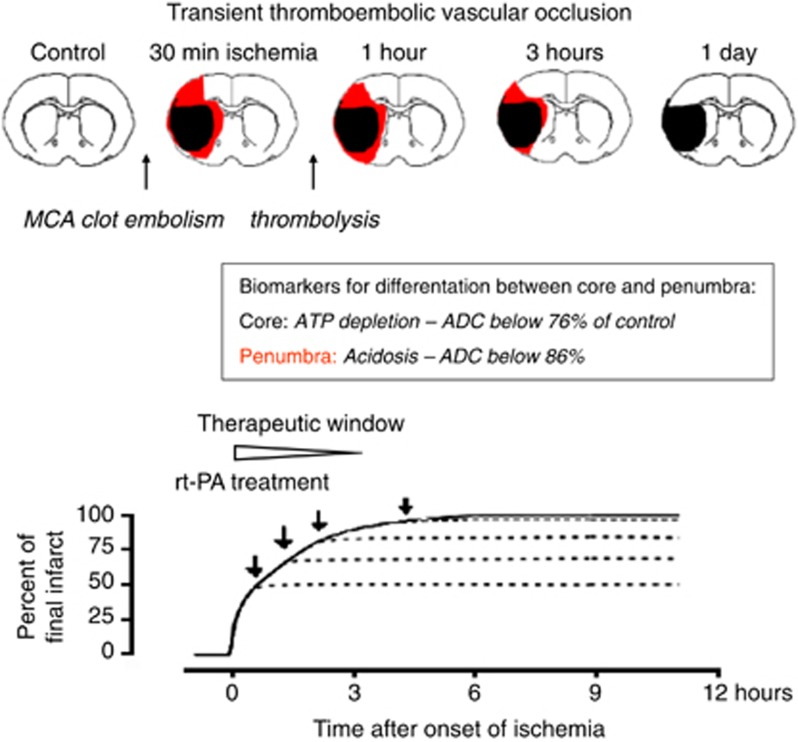
Brain injury after focal ischemia evolves along two basically different pathophysiologies, depending on the severity of the primary flow reduction and the dynamics of postischemic recirculation. In permanent and gradually reversed focal ischemia as after thromboembolic occlusion, primary core injury is irreversible but the expansion of the core into the penumbra can be alleviated by hemodynamic and molecular interventions. Such alleviation can only be achieved within 3 hours after the onset of ischemia because untreated core injury expands to near maximum size during this interval. In promptly reversed transient ischemia as after mechanical vascular occlusion, primary core injury may recover but a secondary delayed injury evolves after a free interval of as long as 6 to 12 hours. This injury can be alleviated throughout the free interval but the longer window is without clinical relevance because transient mechanical vascular occlusion is not a model of naturally occurring stroke. As this difference is widely ignored in stroke research, most clinical trials have been designed with a far too long therapeutic window, which explains their failure. Transient mechanical vascular occlusion models should, therefore, be eliminated from the repertoire of preclinical stroke research.


Figures

Figure 1
Figure 1
Schematic representation of infarct evolution during permanent middle cerebral artery (MCA) occlusion. Core and penumbra are differentiated by biochemical and magnetic resonance imaging according to established viability thresholds of brain ischemia. With ongoing ischemia, the core expands into the penumbra until—within ∼3 hours—infarct reaches its maximum volume. This time is the therapeutic window during which infarct expansion, and hence final infarct volume, can be alleviated.
Figure 2
Figure 2
Dynamics of postischemic recirculation after transient mechanical (A) and recombinant tissue plasminogen activator (rtPA)-treated thromboembolic occlusion (B). Reversal of mechanical occlusion results in prompt recirculation followed by postischemic hyperemia. Recirculation initiated by thrombolysis of clot embolism, in contrast, is only gradually restored and returns to normal after much longer intervals. The different hemodynamics of the two types of ischemia explain that core injury is only reversed after transient mechanical occlusion but not after thrombolytic reperfusion (see Figures 3 and 4; data from Kilic et al, 1999).
Figure 3
Figure 3
Infarct evolution after gradually reversed transient ischemia. Schematic representation of middle cerebral artery (MCA) clot embolism followed by intraarterial infusion of recombinant tissue plasminogen activator (rtPA). Thrombolytic reperfusion does not reverse metabolic failure in the infarct core but it alleviates infarct expansion by improving penumbral blood flow. The therapeutic window is a function of the infarct expansion before the initiation of thrombolysis and is limited to ∼3 hours, as in permanent focal ischemia (see Figure 1).
Figure 4
Figure 4
Infarct evolution after promptly reversed transient ischemia. Schematic representation of increasing durations of middle cerebral artery (MCA) intraluminal filament insertion. The instantaneous reversal of blood circulation after withdrawal of the filament results in prompt reoxygenation of the ischemic tissue and, depending on the duration of ischemia, promotes partial or even complete recovery of energy metabolism in the infarct core. After a free interval of up to 6 to 12 hours secondary delayed cell death evolves in the area of primary energy failure. Secondary delayed cell death can be alleviated by a multitude of molecular interventions throughout the interval between primary and secondary damage. The therapeutic window is, therefore, much longer than after permanent or gradually reversed vascular occlusion. However, this extension is of limited clinical relevance because promptly reversed focal ischemia is not a model of naturally occurring stroke.

References

    1. Astrup J, Symon L, Siesjö BK. Thresholds in cerebral ischemia—The ischemic penumbra. Stroke. 1981;12:723–725. - PubMed
    1. Bath PMW, Gray LJ, Bath AJG, Buchan A, Miyata T, Green AR. Effects of NXY-059 in experimental stroke: an individual animal meta-analysis. Br J Pharmacol. 2009;157:1157–1171. - PMC - PubMed
    1. Busch E, Krüger K, Allegrini PR, Kerskens CM, Gyngell ML, Hoehn-Berlage M, Hossmann K-A. Reperfusion after thrombolytic therapy of embolic stroke in the rat: magnetic resonance and biochemical imaging. J Cereb Blood Flow Metab. 1998;18:407–418. - PubMed
    1. Dirnagl U, Macleod MR. Stroke research at a road block: the streets from adversity should be paved with meta-analysis and good laboratory practice. Br J Pharmacol. 2009;157:1154–1156. - PMC - PubMed
    1. Doyle KP, Simon RP, Stenzel-Poore MP. Mechanisms of ischemic brain damage. Neuropharmacology. 2008;55:310–318. - PMC - PubMed