. 2012 Jan 10;5(206):ra3.
doi: 10.1126/scisignal.2002274.

Wnt/β-catenin signaling and AXIN1 regulate apoptosis triggered by inhibition of the mutant kinase BRAFV600E in human melanoma

Affiliations

Wnt/β-catenin signaling and AXIN1 regulate apoptosis triggered by inhibition of the mutant kinase BRAFV600E in human melanoma

Travis L Biechele et al. Sci Signal. .

Abstract

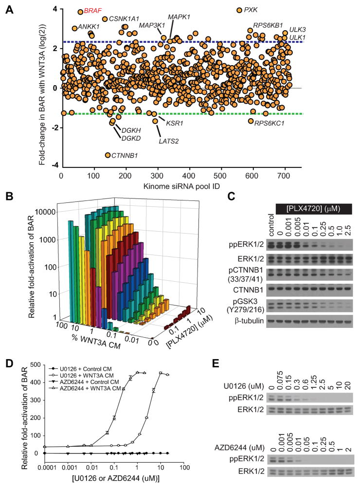
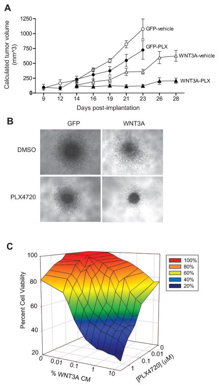
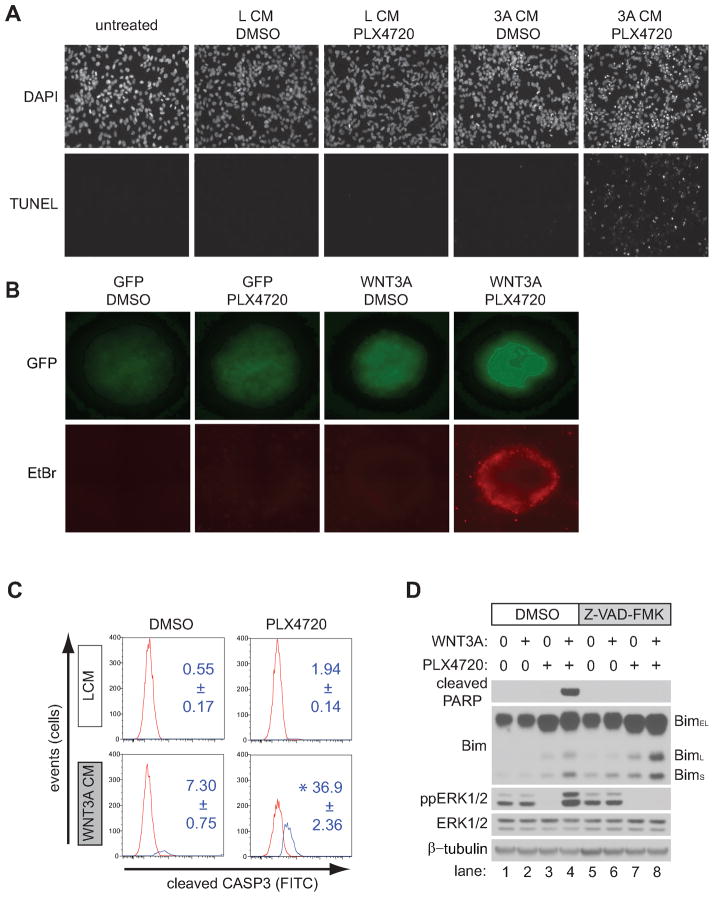
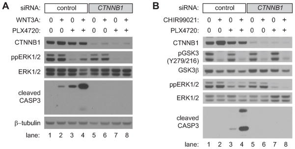
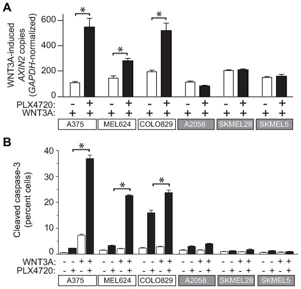
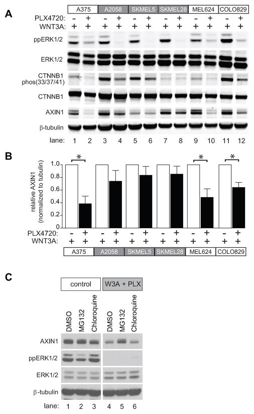
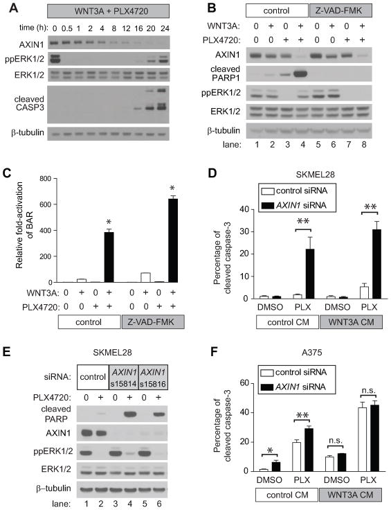
Because the Wnt/β-catenin signaling pathway is linked to melanoma pathogenesis and to patient survival, we conducted a kinome small interfering RNA (siRNA) screen in melanoma cells to expand our understanding of the kinases that regulate this pathway. We found that BRAF signaling, which is constitutively activated in many melanomas by the BRAF(V600E) mutation, inhibits Wnt/β-catenin signaling in human melanoma cells. Because inhibitors of BRAF(V600E) show promise in ongoing clinical trials, we investigated whether altering Wnt/β-catenin signaling might enhance the efficacy of the BRAF(V600E) inhibitor PLX4720. We found that endogenous β-catenin was required for PLX4720-induced apoptosis of melanoma cells and that activation of Wnt/β-catenin signaling synergized with PLX4720 to decrease tumor growth in vivo and to increase apoptosis in vitro. This synergistic enhancement of apoptosis correlated with reduced abundance of an endogenous negative regulator of β-catenin, AXIN1. In support of the hypothesis that AXIN1 is a mediator rather than a marker of apoptosis, siRNA directed against AXIN1 rendered resistant melanoma cell lines susceptible to apoptosis in response to treatment with a BRAF(V600E) inhibitor. Thus, Wnt/β-catenin signaling and AXIN1 may regulate the efficacy of inhibitors of BRAF(V600E), suggesting that manipulation of the Wnt/β-catenin pathway could be combined with BRAF inhibitors to treat melanoma.


Conflict of interest statement

Conflicts of interest: None declared

Competing interests: The authors declare that they have no competing interests.

Figures

Figure 1
Figure 1. BRAF signaling negatively regulates Wnt/β-catenin signaling in melanoma cells
A) Scatter plot of a kinome-based siRNA screen in human A375 melanoma cells stably expressing the β-catenin-activated reporter (BAR) driving firefly luciferase, with each dot representing a known or predicted kinase. Blue- and green-dotted lines represent two mean absolute deviations above and below the mean, respectively. The full gene list is presented in Supplementary database S1–S2. B) An isobologram analysis shows a dose-dependent enhancement of Wnt/β-catenin signaling with the targeted BRAF inhibitor PLX4720 and WNT3A CM on BAR activity. C) Immunoblot analysis of the dose-dependent inhibition of dual-phosphorylated ERK1/2 (ppERK1/2), phosphorylated Ser33/Ser37/Thr41 β-catenin (pCTNNB1), and phosphorylated Tyr216 GSK3 following PLX4720 treatment. D) Two distinct MEK inhibitors, U0126 and AZD6244, both enhanced Wnt/β-catenin signaling in a dose-dependent manner. Symbols and error bars represent the mean and standard deviation, respectively, of three biologic replicates E) Immunoblot analysis of the dose-dependent inhibition of ppERK1/2 by MEK1/2 inhibitors U0126 and AZD6244. In (B–E), A375 cells were treated for 24 hours with the indicated conditions prior to harvesting and data are representative of at least three independent experiments.
Figure 2
Figure 2. Wnt/β-catenin activation cooperates with targeted inhibition of mutant BRAF to inhibit tumor growth in vivo and in vitro
A) WNT3A enhances the ability of the BRAF inhibitor, PLX4720, to reduce tumor size in vivo. Human A375 melanoma cells expressing either GFP or WNT3A(iresGFP) were grown as xenografts in NSG mice treated with either vehicle or 50 mg/kg PLX4720 after tumors had reached an initial size of 100mm3. For each treatment arm, the means and SEM are shown for five individual mice. B) WNT3A enhances the ability of the BRAF inhibitor PLX4720 to reduce spheroid size and in vitro. Human A375 melanoma cells expressing either GFP or WNT3A(iresGFP) were grown as spheroids in a three-dimensional collagen matrix, then treated with either DMSO or 2μM PLX4720 for 72 hours prior to imaging. Representative spheroids of greater than forty spheroids per treatment are shown in these light micrographs. C) WNT3A synergizes with PLX4720 to inhibit the viability of A375 melanoma cells. A375 melanoma cells were treated with the indicated combinations of WNT3A CM and PLX4720 concentrations for 48 hours. In (A–C), data are representative of at least three independent experiments with each data point assayed in triplicate.
Figure 3
Figure 3. Wnt/β-catenin activation synergistically enhances apoptosis with BRAF inhibition
A) TUNEL staining was used to visualize apoptosis in A375 cells following treatment with the indicated conditions, including WNT3A CM (3A CM) or L CM. B) Spheroids generated from A375 cells expressing either GFP or WNT3A(iresGFP) were grown in a three-dimensional collagen matrix and treated with the indicated conditions. Simultaneously, GFP was used to image all cells while EtBr staining was used to identify dead cells. Representative spheroids of greater than forty spheroids for each condition are shown in these panels. C) A flow cytometry-based assay for cleaved caspase-3 was used to detect apoptotic cells following treatment with the indicated conditions in A375 cells. Red and blue peaks on the representative histograms indicate the distribution of cells that were negative and positive for caspase-3 staining respectively. Numbers indicate the average percentage and standard deviation of caspase-3 positive cells from three biological replicates. D) Immunoblot analysis of the proapoptotic protein Bim in A375 cells treated with the indicated conditions. BimEL, BimL, and BimS represent the three major isoforms of Bim. In (A–D), cells were treated for 24 hours with the indicated conditions and data are representative of at least three independent experiments. Where indicated, cells were treated with 2μM PLX4720 and 100μM Z-VAD-FMK.
Figure 4
Figure 4. Apoptosis mediated by Wnt/β-catenin signaling and BRAF inhibition requires β-catenin
A) An immunoblot analysis of cleaved caspase-3 from A375 cells pretreated with control or β-catenin (CTNNB1) siRNA followed by treatment with the indicated conditions. Cells were transfected with siRNAs, incubated for 48 hours, and then treated with the indicated conditions for 48 hours. Where indicated, cells were treated with 2μM PLX4720. B) Immunoblot analysis of cleaved caspase-3 from A375 cells pretreated with control or β-catenin (CTNNB1) siRNA followed by treatment with the indicated conditions. Cells were transfected with siRNAs, incubated for 48 hours, and then treated with the indicated conditions for 36 hours. Where indicated, cells were treated with 2μM PLX4720 and 5μM CHIR99021. Inhibition of GSK3 was confirmed by loss of the activating auto-phosphorylation at Tyr216. In (A–B), data are representative of at least three independent experiments.
Figure 5
Figure 5. Regulation of Wnt/β-catenin signaling by BRAF predicts apoptotic response to combined Wnt/β-catenin activation and BRAF inhibition
A) In six melanoma lines harboring BRAFV600E mutations, synergistic enhancement of Wnt/β-catenin signaling by BRAFV600E inhibition was examined by quantitative PCR measurements of the endogenous target gene AXIN2. Data are expressed as copies of AXIN2 per 106 copies of GAPDH. B) Flow cytometry detection of cleaved caspase-3 was used to measure apoptosis in several melanoma cell lines harboring BRAFV600E mutations. In (A–B), cells were treated with the indicated conditions for 24 hours and 2μM PLX4720 was used where indicated. In (A–B) Columns and error bars represent the mean and standard deviation, respectively, of three biologic replicates. Asterixes (*) represent p<0.001 by one-way ANOVA with Tukey’s post-test. Data are representative of at least three independent experiments.
Figure 6
Figure 6. AXIN1 abundance is positively regulated by BRAF signaling and reduction following BRAF inhibition predicts apoptotic response
A) Immunoblot analysis of the same six melanoma cell lines analyzed in Figure 5 following treatment with the indicated conditions. B) The immunobimmlot of AXIN1 from (A) combined with immunoblots from two additional independent replicates were quantified by pixel intensity. In (A–B), A375 cells were treated with the indicated conditions for 24 hours. C) Reduced AXIN1 abundance following combined treatment with WNT3A and PLX4720 was rescued by the proteasome inhibitor MG132. A375 cells were treated with control conditions or WNT3A and PLX4720 in combination with DMSO or the proteasome inhibitor, MG132 or the lysosome inhibitor, chloroquine for 8 hours. In (A–C) 2μM PLX4720, 10μM MG132, and 10μM chloroquine were used where indicated. In (B) p-values were determined by two-tailed Student’s t-test and (*) represents p<0.005. In (A–C) data are representative of at least three independent experiments.
Figure 7
Figure 7. AXIN1 depletion sensitizes melanoma cells to apoptosis mediated by BRAF inhibition
A) Immunoblot analysis of a time-course of A375 cells treated with WNT3A and PLX4720. B) The decrease in AXIN1 abundance following WNT3A and PLX4720 treatment is not rescued by Z-VAD-FMK. A375 cells were treated for 24 hours with the indicated conditions. C) PLX4720 enhancement of Wnt/β-catenin signaling is not dependent on caspase activation. A375 cells containing the BAR reporter were treated for 24 hours with the indicated conditions. The (*) represents p<0.001 when compared to all other conditions by one-way ANOVA with Tukey’s post-test. D) Flow cytometry detection of cleaved caspase-3 in SKMEL28 cells transfected with control or AXIN1 siRNA and treated with the indicated conditions for 24 hours. E) Knockdown of AXIN1 by siRNA sensitizes SKMEL28 cells to PLX4720-induced apoptosis. Immunoblots show lysates from SKMEL28 cells transfected with either control or two non-overlapping independent siRNAs targeting AXIN1 and treated with the indicated conditions for 24 hours. F) Flow cytometry detection of cleaved caspase-3 in A375 cells transfected with control or AXIN1 siRNA and treated with the indicated conditions for 24 hours. In (D) and (F), p-values were determined by two-way ANOVA with Bonferroni post-test; (*) represents p<0.05 and (**) represents p<0.001. In (A–F) 2μM PLX4720, 10μM MG132, and 100μM Z-VAD-FMK were used where indicated, and data are representative of at least three independent experiments.

Comment in

References

    1. Davies H, Bignell GR, Cox C, Stephens P, Edkins S, Clegg S, Teague J, Woffendin H, Garnett MJ, Bottomley W, Davis N, Dicks E, Ewing R, Floyd Y, Gray K, Hall S, Hawes R, Hughes J, Kosmidou V, Menzies A, Mould C, Parker A, Stevens C, Watt S, Hooper S, Wilson R, Jayatilake H, Gusterson BA, Cooper C, Shipley J, Hargrave D, Pritchard-Jones K, Maitland N, Chenevix-Trench G, Riggins GJ, Bigner DD, Palmieri G, Cossu A, Flanagan A, Nicholson A, Ho JW, Leung SY, Yuen ST, Weber BL, Seigler HF, Darrow TL, Paterson H, Marais R, Marshall CJ, Wooster R, Stratton MR, Futreal PA. Mutations of the BRAF gene in human cancer. Nature. 2002;417:949. doi: 10.1038/nature00766. published online EpubJun 27. nature00766 [pii]) - DOI - PubMed
    1. Tsai J, Lee JT, Wang W, Zhang J, Cho H, Mamo S, Bremer R, Gillette S, Kong J, Haass NK, Sproesser K, Li L, Smalley KS, Fong D, Zhu YL, Marimuthu A, Nguyen H, Lam B, Liu J, Cheung I, Rice J, Suzuki Y, Luu C, Settachatgul C, Shellooe R, Cantwell J, Kim SH, Schlessinger J, Zhang KY, West BL, Powell B, Habets G, Zhang C, Ibrahim PN, Hirth P, Artis DR, Herlyn M, Bollag G. Discovery of a selective inhibitor ofoncogenic B-Raf kinase with potent antimelanoma activity. Proc Natl Acad Sci U S A. 2008;105:3041. doi: 10.1073/pnas.0711741105. published online EpubFeb 26. 0711741105 [pii] - DOI - PMC - PubMed
    1. Bollag G, Hirth P, Tsai J, Zhang J, Ibrahim PN, Cho H, Spevak W, Zhang C, Zhang Y, Habets G, Burton EA, Wong B, Tsang G, West BL, Powell B, Shellooe R, Marimuthu A, Nguyen H, Zhang KY, Artis DR, Schlessinger J, Su F, Higgins B, Iyer R, D’Andrea K, Koehler A, Stumm M, Lin PS, Lee RJ, Grippo J, Puzanov I, Kim KB, Ribas A, McArthur GA, Sosman JA, Chapman PB, Flaherty KT, Xu X, Nathanson KL, Nolop K. Clinical efficacy of a RAF inhibitor needs broad target blockade in BRAF-mutant melanoma. Nature. 2010;467:596. doi: 10.1038/nature09454. published online EpubSep 30. nature09454 [pii] - DOI - PMC - PubMed
    1. Lee JT, Li L, Brafford PA, van den Eijnden M, Halloran MB, Sproesser K, Haass NK, Smalley KS, Tsai J, Bollag G, Herlyn M. PLX4032, a potent inhibitor of the B-Raf V600Eoncogene, selectively inhibits V600E-positive melanomas. Pigment Cell Melanoma Res. 2010;23:820. doi: 10.1111/j.1755-148X.2010.00763.x. published online EpubDec (PCR763 [pii] - DOI - PMC - PubMed
    1. Kefford RF, Arkenau H, Brown MP, Millward M, Infante JR, Long GV, Ouellet D, Curtis M, Lebowitz PF, Falchook GS. Phase I/II study of GSK2118436, a selective inhibitor of oncogenic mutant BRAF kinase, in patients with metastatic melanoma and other solid tumors. J Clin Oncol. 2010;28:abstract 8503. published online Epub2010 (

Publication types