. 2012 Mar 1;119(9):1992-2002.
doi: 10.1182/blood-2011-06-354647. Epub 2012 Jan 10.

p38 MAPK inhibition suppresses the TLR-hypersensitive phenotype in FANCC- and FANCA-deficient mononuclear phagocytes

Affiliations

p38 MAPK inhibition suppresses the TLR-hypersensitive phenotype in FANCC- and FANCA-deficient mononuclear phagocytes

Praveen Anur et al. Blood. .

Abstract

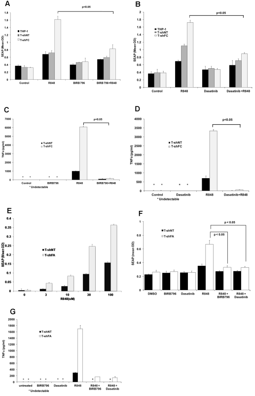
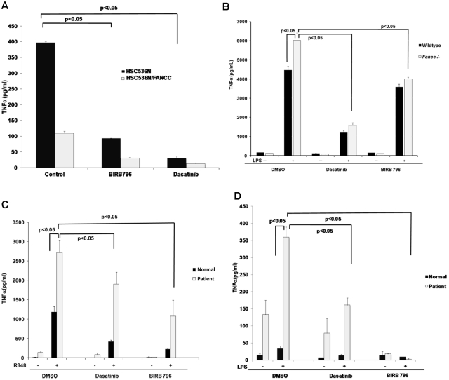
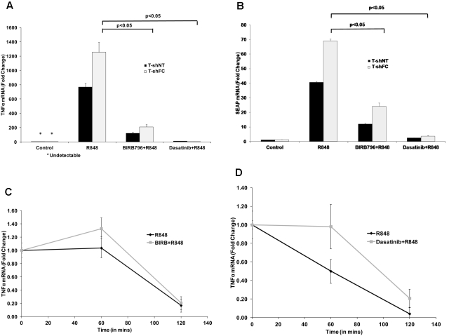
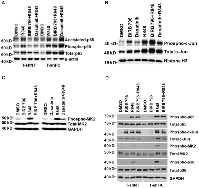
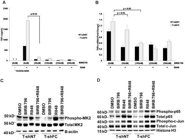
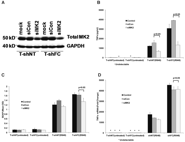
Fanconi anemia, complementation group C (FANCC)-deficient hematopoietic stem and progenitor cells are hypersensitive to a variety of inhibitory cytokines, one of which, TNFα, can induce BM failure and clonal evolution in Fancc-deficient mice. FANCC-deficient macrophages are also hypersensitive to TLR activation and produce TNFα in an unrestrained fashion. Reasoning that suppression of inhibitory cytokine production might enhance hematopoiesis, we screened small molecules using TLR agonist-stimulated FANCC- and Fanconi anemia, complementation group A (FANCA)-deficient macrophages containing an NF-κB/AP-1-responsive reporter gene (SEAP). Of the 75 small molecules screened, the p38 MAPK inhibitor BIRB 796 and dasatinib potently suppressed TLR8-dependent expression of the reporter gene. Fanconi anemia (FA) macrophages were hypersensitive to the TLR7/8 activator R848, overproducing SEAP and TNFα in response to all doses of the agonist. Low doses (50nM) of both agents inhibited p38 MAPK-dependent activation of MAPKAPK2 (MK2) and suppressed MK2-dependent TNFα production without substantially influencing TNFα gene transcription. Overproduction of TNFα by primary FA cells was likewise suppressed by these agents and involved inhibition of MK2 activation. Because MK2 is also known to influence production and/or sensitivity to 2 other suppressive factors (MIP-1α and IFNγ) to which FA hematopoietic progenitor cells are uniquely vulnerable, targeting of p38 MAPK in FA hematopoietic cells is a rational objective for preclinical evaluation.


Figures

Figure 1
Figure 1
BIRB 796 and dasatinib inhibit NF-κB/AP-1–dependent SEAP production in T-shFC cells. Mean values ± SD are shown. All P values were calculated using the Student t test. (A) BIRB 796 inhibits R848-induced SEAP gene expression in FANCC-deficient cells. THP1-Blue cells (THP1) or THP1-Blue cells stably expressing either control (nontarget) shRNA (T-shNT) or shRNA targeting FANCC (T-shFC) were cultured in medium alone (control) or medium with the TLR7/8 agonist R848 (30μM) alone, BIRB 796 (500nM) alone, or R848 (30μM) alone after a 6-hour pretreatment with BIRB 796 (500nM). Conditioned media were collected after 24 hours of R848 exposure and SEAP was quantified colorimetrically using QUANTI-Blue reagent. (B) Dasatinib inhibits R848-induced SEAP expression in FANCC-deficient cells. The experimental design was identical to that of 1A except that dasatinib (500nM) was used as the inhibitor. (C) BIRB 796 inhibits TNFα secretion in THP-1 cells. T-shNT and T-shFC were treated as in panel A with BIRB 796 (500nM) and/or R848 (30μM). Conditioned media were collected after 24 hours of incubation with R848 and TNFα was quantified by ELISA. Untreated cells or cells treated only with BIRB 796 did not produce detectable TNFα (indicated with an asterisk). (D) Dasatinib inhibits TNFα secretion in THP-1 cells. T-shNT and T-shFC cells were treated as in panel B with R848 (30μM) and dasatinib (500nM). Untreated cells or cells treated with dasatinib alone did not produce detectable TNFα (indicated with an asterisk). (E) FANCA-deficient THP-1 cells are hypersensitive to R848. THP1-Blue cells stably expressing shRNA targeting FANCA (T-shFA) and T-shNT cells were exposed to R848 (0-100μM) for 24 hours. Supernatant media were collected and SEAP was quantified colorimetrically using QUANTI-Blue reagent. At each dose of R848, T-shFA cells produced more SEAP than did T-shNT cells (P < .05). (F) BIRB 796 and dasatinib suppress the production of SEAP in both T-shFA and T-shNT cells. The design of these studies on T-shFA cells was identical to that of studies on T-shFC cells (A-B). (G) BIRB 796 and dasatinib suppress TNFα production in R848-stimulated T-shFA and T-shNT cells. The design of these studies on T-shFA cells was identical to that used with T-shFC cells (C-D).
Figure 2
Figure 2
BIRB 796 and dasatinib suppress TLR-activated TNFα gene expression in primary and patient-derived FA cells. Means ± SD are shown. All P values were calculated using the Student t test. (A) BIRB 796 and dasatinib inhibit constitutive TNFα secretion in the patient-derived FANCC−/− lymphoblast cell line HSC536N. HSC536N and HSC536N/FANCC (complemented with normal FANCC cDNA) were cultured in medium alone (control) or medium treated with BIRB 796 (500nM) or dasatinib (500nM) for 6 hours. After 6 hours, supernatant media were collected and TNFα measured by ELISA. (B) BIRB 796 and dasatinib inhibit LPS-induced TNFα production in Fancc-deficient murine macrophages. Primary murine BM-derived macrophages obtained from Fancc−/− and wild-type mice were maintained in control medium (plus DMSO) or treated for 6 hours with BIRB 796 (500nM) or dasatinib (500nM), followed by stimulation with LPS (1 μg/mL) for 24 hours. TNFα in conditioned media was measured by ELISA. (C-D) BIRB 796 and dasatinib inhibit R848- and LPS-induced TNFα production in FANCA-deficient human macrophages. Primary human CD14+ macrophages obtained from a normal age-matched volunteer and a FANCA-deficient patient were cultured for 6 hours in control medium (plus DMSO), BIRB 796 (500nM), or dasatinib (500nM), before stimulation for 24 hours with R848 (1μM; C) or LPS (0.1 ng/mL; D). TNFα in conditioned media was measured by ELISA.
Figure 3
Figure 3
BIRB 796 and dasatinib suppress the production of TNFα and SEAP transcripts. T-shNT and T-shFC were maintained in medium alone (control), or medium treated with R848 (30μM) for 24 hours after 6 hours of pretreatment with either BIRB 796 (500nM) or dasatinib (500nM). Total RNA was prepared and TNFα (A) and SEAP (B) mRNAs quantified using real-time qRT-PCR. Data are expressed as the mean ± SD -fold change relative to transcripts in unexposed (control) cells and based on 3 technical replicates normalized to 18S rRNA. Dasatinib suppressed TNFα mRNA levels to a greater degree than did BIRB 796. (C-D) Neither BIRB 796 nor dasatinib enhanced TNFα mRNA degradation. T-shFC cells were either cultured in medium alone or treated with BIRB 796 (500nM; C) or dasatinib (500nM; D) for 6 hours, after which time all cells were stimulated with R848 (30μM) for 24 hours. After adding actinomycin D (5 μg/mL), total RNA was collected at 0, 60, and 120 minutes and TNFα mRNA was measured by real-time qRT-PCR. For each drug, 1 representative of 2 experiments with identical results is shown.
Figure 4
Figure 4
Dasatinib and BIRB 796 do not inhibit R848-induced NF-κB p65 or c-Jun levels in FANCC- or FANCA-deficient mononuclear phagocytes, but do inhibit MK2 phosphorylation. T-shNT cells and T-shFC cells were treated with BIRB 796 (500nM) or dasatinib (500nM) for 6 hours and then stimulated with R848 (30μM) for 24 hours. (A) Western blot analyses of nuclear extracts were performed using Abs for total p65, phospho-p65(Ser536), and acetylated p65(Lys310). Whereas both BIRB 796 and dasatinib suppressed p65 phosphorylation and acetylation in T-shNT cells, neither agent suppressed p65 phosphorylation in T-shFC cells, and only BIRB 796 inhibited p65 acetylation. One representative example of 3 identical experiments is shown. (B) Western blot analyses of nuclear extracts were performed as above, but Abs to total c-Jun and phosphorylated (Ser73) c-Jun were used. BIRB 796 did not suppress c-Jun phosphorylation and the suppressive effect of dasatinib was minimal (lane 6 vs lane 4). One representative example of 3 identical experiments is shown. (C) Immunoblot analysis of whole-cell lysates of shFC cells exposed to R848 with and without either dasatinib or BIRB 796 demonstrated marked suppression of MK2 phosphorylation by both small molecules (lanes 4 and 8 vs lanes 3 and 7). One representative example of 3 identical experiments is shown. (D) T-shFA and T-shNT cells were exposed to R848 alone for 24 hours or to BIRB 796 for 6 hours, followed by R848 for 24 hours. Whole-cell lysates were used in immunoblot assays for total and phosphorylated p65, c-jun, p38, and MK2. BIRB 796 had no influence on p65 or c-jun phosphorylation, but profoundly suppressed p38 and MK2 phosphorylation. One representative experiment of 2 is shown.
Figure 5
Figure 5
BIRB 796 and dasatinib influence TNFα gene expression posttranscriptionally. Means ± SD are shown. All P values were calculated using the Student t test. (A) BIRB 796 effectively inhibited secreted TNFα in monocytes. T-shNT and T-shFC cells were treated with BIRB 796 (0-500nM) for 6 hours, followed by treatment with R848 (30μM) for 24 hours. Supernatant media were then collected and TNFα was quantified by ELISA. Low doses of BIRB 796 (50nM) suppressed secreted TNFα protein by 20-fold. (B) T-shNT and T-shFC cells were treated with BIRB 796 (0-500nM) for 6 hours, followed by treatment with R848 (30μM) for 24 hours. mRNA was then prepared and TNFα transcripts measured by real-time qRT-PCR. Data are expressed as the fold change normalized to 18S rRNA and are relative to mRNA levels in unexposed (control) T-shNT cells. TNFα mRNA is reduced modestly in the presence of BIRB 796 and minimally at the 50nM dose that was fully suppressive of TNFα protein production (A). One of 2 identical experiments is shown. (C) Low doses of BIRB 796 (50nM) suppress R848-induced MK2 phosphorylation in whole-cell lysates from T-shFC and T-shNT cells. One of 2 identical experiments is shown. (D) Immunoblot analysis for p65 and c-Jun phosphorylation in nuclear extracts of T-shFC and T-shNT cells revealed that low doses of BIRB 796 had no effect on p65 or c-Jun phosphorylation in R848-induced T-shFC and T-shNT cells. One representative immunoblot of 3 identical experiments is shown.
Figure 6
Figure 6
MK2 regulates TNFα production posttranscriptionally. T-shFC and T-shNT cells were transfected with nontargeted siRNAs (siCon) or siRNA targeting MK2 (siMK2) by nucleofection. Cells were also subjected to nucleofection without siRNA present (mock). At the time of maximal MK2 knockdown (72 hours), the cells were stimulated with R848 (30μM) for 24 hours or were left untreated. (A) MK2 suppression (lanes 3 and 6) was confirmed by Western blot of whole-cell extracts 72 hours after nucleofection. (B) TNFα protein in supernatant media was quantified by ELISA 24 hours after R848 stimulation. siMK2 suppressed TNFα levels produced by both T-shFC and T-shNT cells and the degree of suppression matched the degree of MK2 knockdown with RNAi (6A). (C) SEAP expression, as quantified by QUANTI-Blue, was minimally suppressed by siMK2. (D) TNFα mRNA measured 24 hours after R848 stimulation by real-time qRT-PCR revealed no suppression by treatment of cells with siMK2. Therefore, MK2 suppression inhibited TNFα production and/or release, but not SEAP or TNFα gene transcription in T-shFC cells.

References

    1. de Winter JP, Joenje H. The genetic and molecular basis of Fanconi anemia. Mutat Res. 2009;668(1-2):11–19. - PubMed
    1. Kim Y, Lach FP, Desetty R, Hanenberg H, Auerbach AD, Smogorzewska A. Mutations of the SLX4 gene in Fanconi anemia. Nat Genet. 2011;43(2):142–146. - PMC - PubMed
    1. Stoepker C, Hain K, Schuster B, et al. SLX4, a coordinator of structure-specific endonucleases, is mutated in a new Fanconi anemia subtype. Nat Genet. 2011;43(2):138–141. - PubMed
    1. Vaz F, Hanenberg H, Schuster B, et al. Mutation of the RAD51C gene in a Fanconi anemia-like disorder. Nat Genet. 2010;42(5):406–409. - PubMed
    1. Hays LE, Keeble WW, Yates JE, et al. Human FANCC is hypomorphic in murine Fancc-deficient cells. Blood. 2010;116(12):2057–2060. - PMC - PubMed

Publication types

MeSH terms