. 2012 May;40(9):3898-912.
doi: 10.1093/nar/gkr1296. Epub 2012 Jan 10.

Structural mechanism of the phosphorylation-dependent dimerization of the MDC1 forkhead-associated domain

Affiliations

Structural mechanism of the phosphorylation-dependent dimerization of the MDC1 forkhead-associated domain

Jinping Liu et al. Nucleic Acids Res. 2012 May.

Abstract

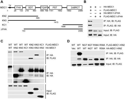
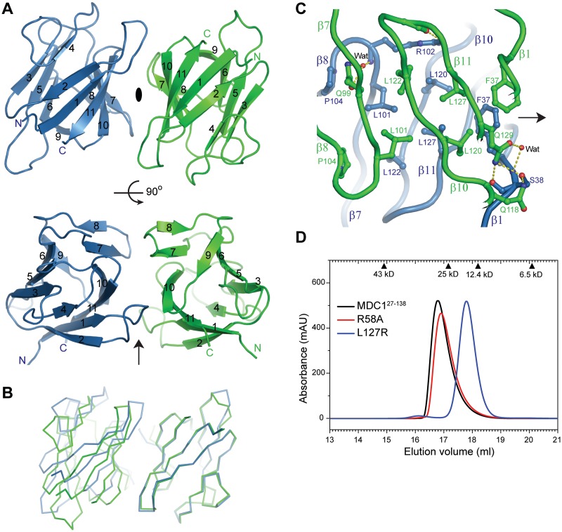
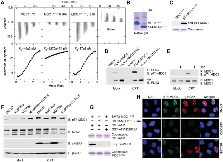
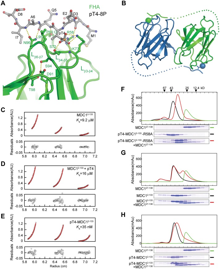
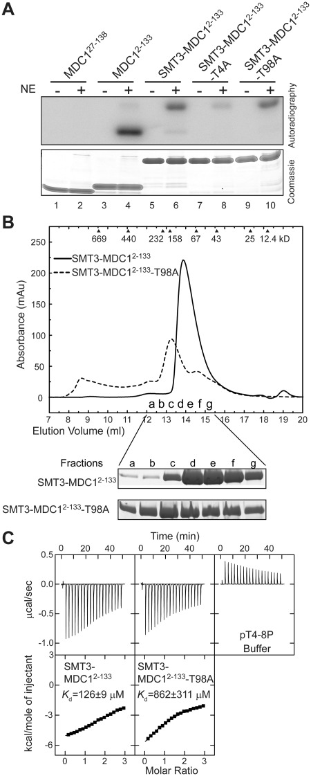
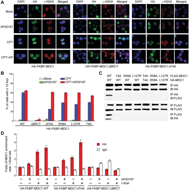
MDC1 is a key mediator of the DNA-damage response in mammals with several phosphorylation-dependent protein interaction domains. The function of its N-terminal forkhead-associated (FHA) domain remains elusive. Here, we show with structural, biochemical and cellular data that the FHA domain mediates phosphorylation-dependent dimerization of MDC1 in response to DNA damage. Crystal structures of the FHA domain reveal a face-to-face dimer with pseudo-dyad symmetry. We found that the FHA domain recognizes phosphothreonine 4 (pT4) at the N-terminus of MDC1 and determined its crystal structure in complex with a pT4 peptide. Biochemical analysis further revealed that in the dimer, the FHA domain binds in trans to pT4 from the other subunit, which greatly stabilizes the otherwise unstable dimer. We show that T4 is phosphorylated primarily by ATM upon DNA damage. MDC1 mutants with the FHA domain deleted or impaired in its ability to dimerize formed fewer foci at DNA-damage sites, but the localization defect was largely rescued by an artificial dimerization module, suggesting that dimerization is the primary function of the MDC1 FHA domain. Our results suggest a novel mechanism for the regulation of MDC1 function through T4 phosphorylation and FHA-mediated dimerization.


Figures

Figure 1.
Figure 1.
MDC1 self-associates via its FHA domain. (A) Diagram of MDC1 domain organization. Boundaries are indicated for MDC1 truncation constructs. (B–D) HA-tagged MDC1 or mutants were co-transfected with FLAG-tagged MDC1 or mutants into Ad293 cells. After 48 h, the total cell lysates were immunoprecipitated with anti-FLAG and/or anti-HA antibodies and immunoblotted with anti-HA and anti-FLAG antibodies. The whole cell lysates were also blotted against anti-HA and -FLAG antibodies and are shown as the input. (B) Self-association of MDC1 and its FHA deletion mutant (MDC1ΔFHA) were analyzed by immunoprecipitation (IP) and immunoblotting (IB). (C) Self-association of wild-type MDC1 (WT) and its truncation mutants KN2, KN3 and KC1. (D) Self-association of KN2 and its mutants R58A and N96A/G97A/T98A (NGT).
Figure 2.
Figure 2.
Dimeric structure of the MDC1 FHA domain. (A) Ribbon representation of the FHA dimer structure in the P212121 space group. Two orthogonal views are displayed. The pseudo-dyad axis is shown as an ellipse when perpendicular to the paper or as an arrow when in the paper. The two subunits are colored green and blue. The 11 strands are labeled by numbers, and the N- and C-termini are marked. (B) Alignment of two dimers by the non-equivalent protomers to the right. The other protomers to the left do not overlap following the alignment, indicating asymmetry in the dimer arrangement. (C) Interactions at the dimer interface. Protein backbones are shown as tubes, interacting residues as sticks and hydrogen bonds as dashed lines. Carbon atoms are green in one subunit and blue in the other subunit, oxygen atoms are red and nitrogen atoms are blue. (D) MDC127–138 and its mutants R58A and L127R were analyzed in a Superdex 200 10/300 GL column running in HEPES–Na (pH 7.6) and 200 mM NaCl. Each protein (400 μM) was loaded in a 100 μl volume. The elution volume of calibration standards are indicated on the top.
Figure 3.
Figure 3.
MDC1 T4 is phosphorylated by ATM in response to DNA damage, and pT4 is recognized by the FHA domain. (A) Microcalorimetric titration of phosphopeptide pT4-8P into MDC127–138, its mutants R58A and L127R, or 20 mM sodium phosphate (pH 7.6) and 150 mM NaCl buffer. The heat peaks were integrated, corrected for the ligand dilution effect and fit to a one-set-of-sites binding model. The dissociation constant, Kd, values are indicated. (B) MDC12–133 was phosphorylated by nuclear extracts (NE) of HeLa cells and resolved in a native gel. (C) Affinity purified rabbit polyclonal anti-pT4-MDC1 antibody specifically recognizes phosphorylated MDC12–133 but not unphosphorylated MDC12–133. (D) pT4 in transiently expressed MDC1. Empty vector or vectors expressing FLAG-MDC1 or FLAG-MDC1(T4A) were transfected into U2OS cells. The cells were mock treated or treated with CPT for 1 h, followed by immunoprecipitation with anti-FLAG antibody and immunoblotting with anti-FLAG and anti-pT4-MDC1 antibodies. (E) pT4 in endogenous MDC1. U2OS cells were mock treated or treated with CPT for 1 h. The total cell lysates were immunoprecipitated with anti-MDC1 antibody, treated with or without calf intestine phosphatase (CIP), and blotted with anti-pT4-MDC1 and anti-MDC1 antibodies. (F) T4 is primarily phosphorylated by ATM. U2OS cells were treated with DMSO, ATM inhibitor KU55993 (10 μM), DNA-PKcs inhibitor NU7026 (10 μM) or both KU55993 and NU7026 for 2 h before treatment of 0 or 10 μM CPT for 1 h. The total cell lysates were resolved in SDS-PAGE and blotted with anti-pT4-MDC1, anti-MDC1, anti-γ-H2AX and anti-β-actin antibodies. Endogenous MDC1 presents multiple isoforms when blotted with anti-MDC1 antibody; only the slowest migrating one was recognized by anti-pT4-MDC1 antibody. (G) SMT3-MDC12–133, but not its T4A mutant, can be phosphorylated in vitro by a recombinant GST-fused kinase domain of ATM (GST-ATM, residues 2709–2964). GST-ATM-D2870A is a kinase-dead mutant. (H) U2OS cells were treated as in (F) and were immunostained with anti-pT4-MDC1 (green) and γ-H2AX (red). DNA was stained by DAPI (blue).
Figure 4.
Figure 4.
MDC FHA domain binds to pT4 in trans. (A) The monomeric structure of MDC127–138 in complex with phosphopeptide pT4-8P. The phosphopeptide and important ligand-binding residues are shown as sticks and balls and colored red for oxygen, blue for nitrogen, gray for carbon in the phosphopeptide and green for carbon in the FHA domain. (B) A structural model depicting the trans FHA–pT4 interaction in the FHA dimer. Dots denote putative positions of residues 9–26. Phosphate groups are shown as spheres. (C–E) Analytic ultracentrifugation sedimentation equilibrium analysis of MDC12–133(C), MDC12–133 in the presence of pT4-8P (D) and pT4-MDC12–133(E). The dimer dissociation constants Kd are indicated. (F–H) Subunit exchange between MDC12–133 and MDC127–138 homodimers. pT4-MDC12–133-R58A (F), MDC12–133 (G) and pT4-MDC12–133 (H) were analyzed by a Superdex 75 10/300 GL column in the absence and presence of MDC127–138. The fractions were resolved with SDS-PAGE, and the gels are shown below the elution profiles. The same elution profile and gel image of MDC127–138 are displayed in each panel for comparison.
Figure 5.
Figure 5.
T98 is not a phosphorylation site and the T98A mutation disturbs the folding and pT-binding of the FHA domain. (A) Phosphorylation of MDC1 FHA proteins by HeLa cell nuclear extracts (NE) in the presence of 32P-γ-ATP. (B) SMT3-MDC12–133 and its T98A mutant were eluted in a Superdex 200 10/300 GL column and in buffer 20 mM sodium phosphate (pH 7.6) and 250 mM NaCl. SDS-PAGE gels of the fractions are shown at the bottom. (C) ITC analysis of SMT3-MDC12–133 and its T98A mutant with phosphopeptide pT4-8P in 20 mM sodium phosphate (pH 7.6) and 250 mM NaCl.
Figure 6.
Figure 6.
Dimerization promotes enrichment of MDC1 at DNA-damage sites. (A) HA-FKBP-MDC1, HA-FKBP-MDC1ΔBRCT or HA-FKBP-MDC1ΔFHA was stably expressed in Ad293 cells. The cells were mock treated or treated with 10 μM CPT for 1 h, with 10 nM AP20187 for 4 h to induce dimerization of FKBP, or with 10 nM AP20187 for 4 h and 10 μM CPT for 1 h. Cells were fixed and immuno-stained with anti-HA (green) and anti-γ-H2AX (red). DNA was stained with DAPI (blue). (B) Percentage of cells with 10 or more HA-foci in (A). Average of three independent experiments ± SD. In each treatment, 100–200 cells were counted. (C) The T4A, R58A or L127R mutations disrupt MDC1 dimerization. HA- and FLAG-tagged MDC1 or the indicated mutants were transiently expressed in Ad293 cells for 48 h. The total cell lysates were immunoprecipitated with anti-HA and anti-FLAG antibodies and immunoblotted with anti-HA and anti-FLAG antibodies. (D) ChIP analysis of MDC1 localization in defined DSBs. HA-FKBP-MDC1, HA-FKBP-MDC1ΔBRCT or HA-FKBP-MDC1ΔFHA was stably expressed in DFGFP U2OS cells. The cells were transfected with or without an I-SceI expression construct for 24 h, treated with 0 or 10 nM AP20187, and harvested for ChIP assays with anti-HA antibody or a control IgG. The input sample prior to immunoprecipitation and the ChIP elutant were amplified by real-time PCR using a pair of primers covering a region 190–335 bp from the I-SceI cutting site. The signal intensity of the HA or IgG ChIP samples was first divided by the signal intensity of the input samples of same treatment and then normalized to the signal intensity of the ChIP samples without AP20187 and I-SceI treatment. Average of three measures ± SD.

Comment on

  • Nucleic Acids Res.

Similar articles

Cited by

References

    1. Ciccia A, Elledge SJ. The DNA damage response: making it safe to play with knives. Mol. Cell. 2010;40:179–204. - PMC - PubMed
    1. Ohnishi T, Mori E, Takahashi A. DNA double-strand breaks: their production, recognition, and repair in eukaryotes. Mutat. Res. 2009;669:8–12. - PubMed
    1. Mahaney BL, Meek K, Lees-Miller SP. Repair of ionizing radiation-induced DNA double-strand breaks by non-homologous end-joining. Biochem. J. 2009;417:639–650. - PMC - PubMed
    1. Williams RS, Williams JS, Tainer JA. Mre11-Rad50-Nbs1 is a keystone complex connecting DNA repair machinery, double-strand break signaling, and the chromatin template. Biochem. Cell Biol. 2007;85:509–520. - PubMed
    1. Rogakou EP, Pilch DR, Orr AH, Ivanova VS, Bonner WM. DNA double-stranded breaks induce histone H2AX phosphorylation on serine 139. J. Biol. Chem. 1998;273:5858–5868. - PubMed

Publication types

MeSH terms

Substances