. 2012 Dec;22(12):2715-32.
doi: 10.1093/cercor/bhr295. Epub 2012 Jan 10.

The left occipitotemporal cortex does not show preferential activity for words

Affiliations

The left occipitotemporal cortex does not show preferential activity for words

Alecia C Vogel et al. Cereb Cortex. 2012 Dec.

Abstract

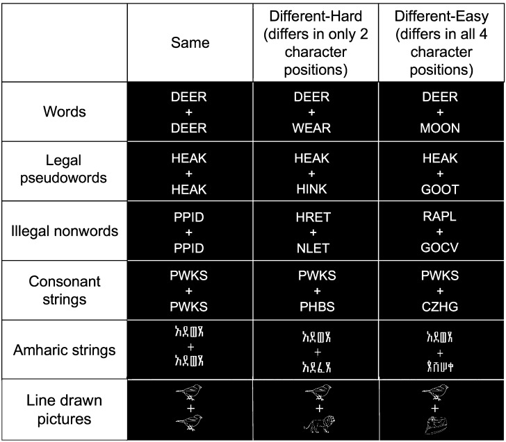
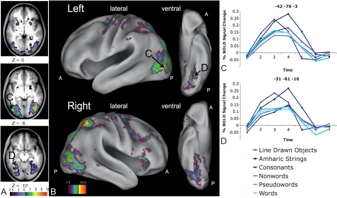
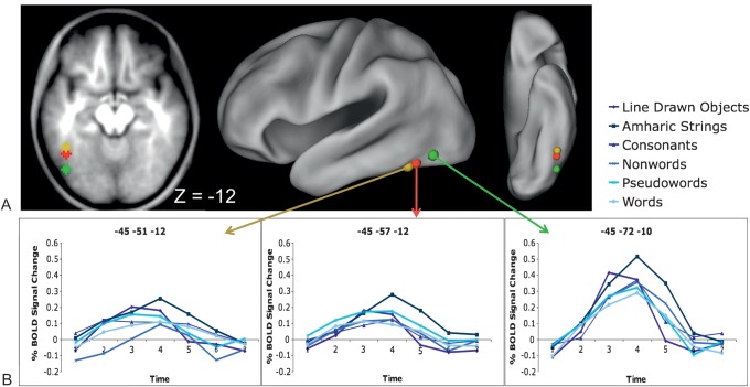
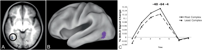
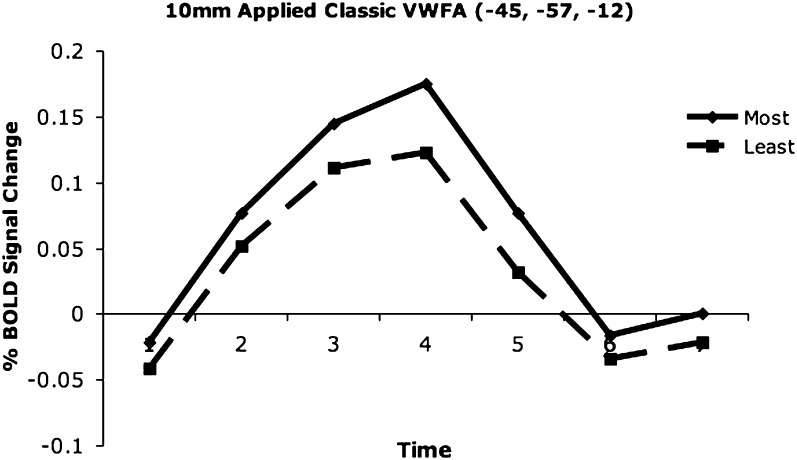
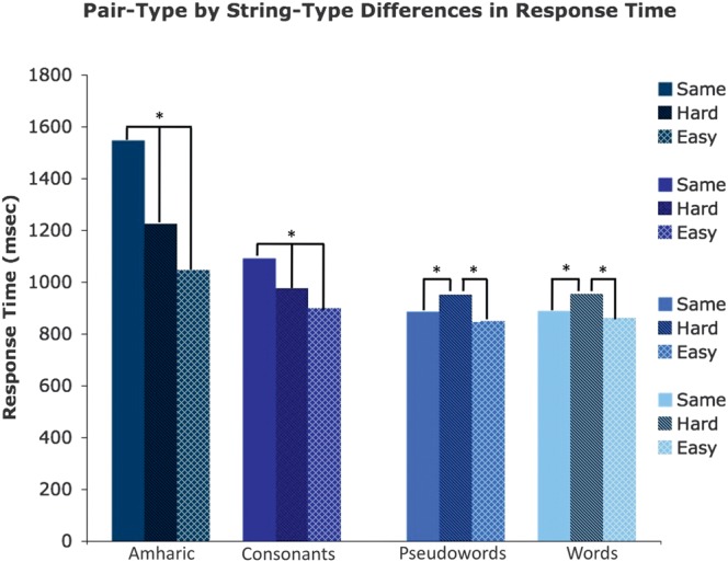
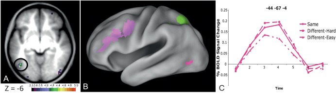
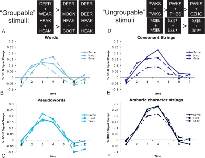
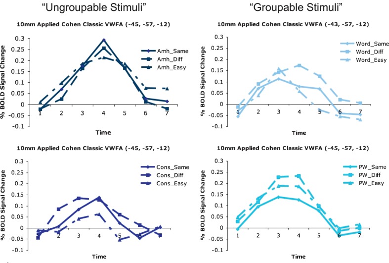
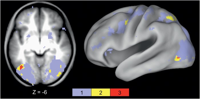
Regions in left occipitotemporal (OT) cortex, including the putative visual word form area, are among the most commonly activated in imaging studies of single-word reading. It remains unclear whether this part of the brain is more precisely characterized as specialized for words and/or letters or contains more general-use visual regions having properties useful for processing word stimuli, among others. In Analysis 1, we found no evidence of greater activity in left OT regions for words or letter strings relative to other high-spatial frequency high-contrast stimuli, including line drawings and Amharic strings (which constitute the Ethiopian writing system). In Analysis 2, we further investigated processing characteristics of OT cortex potentially useful in reading. Analysis 2 showed that a specific part of OT cortex 1) is responsive to visual feature complexity, measured by the number of strokes forming groups of letters or Amharic strings and 2) processes learned combinations of characters, such as those in words and pseudowords, as groups but does not do so in consonant and Amharic strings. Together, these results indicate that while regions of left OT cortex are not specialized for words, at least part of OT cortex has properties particularly useful for processing words and letters.


Figures

Figure 1.
Figure 1.
Examples of stimulus pairs. Six types of stimulus pairs were used in a visual matching judgment task. Fifty percent of the pairs presented were the same (as seen in the second column), 25% were hard, 2-character different pairs (as seen in the third column), and 25% were easy, all different pairs (as seen in the fourth column). In the picture-matching run (bottom row), pictures were drawn from the same semantically related category for the hard condition and from separate semantic categories for the easy condition.
Figure 2
Figure 2
Left fusiform regions showing a stimulus type by timecourse interaction. (A) Image derived from the whole-brain analysis, focusing on slices in occipital and fusiform cortex, showing voxels with a significant stimulus type by timecourse interaction. (B) Whole-brain analysis image showing all voxels with a significant stimulus type by timecourse interaction projected to the surface of semi-inflated brain surfaces rendered with CARET (Van Essen et al. 2001; http://brainmap.wustl.edu/caret). Letter labels indicate regions for which timecourses are shown in panels (C) and (D). (C) Timecourses for all 6 stimulus types in a left fusiform region posterior and superior to the classically described VWFA (−42, −76, −3 in MNI coordinates). Further ANOVAs show this interaction is due to more activity for Amharic characters than pictures (P < 0.001) and letter strings (P < 0.001 for all) and more activity for pictures than letter strings (P < 0.001 for all). (D) Timecourses for all 6 stimulus types in a left fusiform region anterior and medial to the classically described VWFA (−31, −61, −10 in MNI coordinates). Further ANOVAs show this interaction is due to more activity for Amharic characters than pictures (P < 0.001) and letter strings (P < 0.001 for all) and more activity for pictures than letter strings (P < 0.001 for all).
Figure 3.
Figure 3.
Stimulus effects in literature-derived pVWFA regions. (A) Location of applied pVWFA regions from Cohen and Dehaene (2004), displayed on a transverse section through fusiform cortex and on a semi-inflated CARET surface. (B) Timecourses for all 6 stimulus types in the applied VWFA regions. There was no stimulus type by timecourse interaction in the anterior (−45, −51, −12, MNI coordinates) and classic (−45, −57, −12, MNI coordinates) regions. There is a significant stimulus type by timecourse interaction in the posterior (−45, −72, −10, MNI coordinates) region (P < 0.05), which post hoc ANOVAs show is due to trend-level greater activity for Amharic characters than consonants, pseudowords, and words (all P < 0.10) and significantly greater activity for pictures than nonwords, pseudowords, and words (all P < 0.04).
Figure 4.
Figure 4.
Left OT region showing a complexity by timecourse interaction. (A) Location of voxels showing a visual complexity by timecourse interaction Z-score > 3.5 in a transverse slice through fusiform cortex. The circled OT region (−40, −64, −4 in MNI coordinates) was the only left hemisphere region identified. (B) Location of the left OT region from panel (A) showing a visual complexity by timecourse interaction on a semi-inflated CARET surface. (C). Timecourses for the most and least visually complex pairs (all stimulus types averaged together) in the left OT region identified from the whole-brain complexity by timecourse analysis. This region shows more activity for the most complex relative to the least complex pairs (P = 0.013).
Figure 5.
Figure 5.
Complexity timecourses in the applied pVWFA ROI. There is more activity for the most visually complex pairs of stimuli relative to the least complex pairs in an applied, classically defined pVWFA region (Cohen and Dehaene 2004).
Figure 6.
Figure 6.
RT to match various pair types for each stimulus type. RT to match Amharic character and consonant strings increases with the number of characters that must be evaluated to make the matching decision. The RTs are significantly different for all pair types in these stimuli. RTs to match pseudowords and words are equivalent for the same and easy (4-different) pairs, which are faster to match than hard (2-different) pairs. All statistical effects are denoted with asterisks indicating P < 0.05. Error bars indicate the standard error of the mean.
Figure 7.
Figure 7.
Left OT region showing a pair type by timecourse interaction. (A) Location of voxels showing a pair type by timecourse interaction Z-score > 3.5 in a transverse slice through fusiform cortex. The peak of the left OT region is located at −44, −67, −4 in MNI coordinates. (B) All left lateral hemisphere regions showing a pair type by timecourse interaction on a semi-inflated CARET surface. In fuchsia is the OT region shown in panel (A). Pink regions show a similar pattern of effects as the fuchsia region (BOLD activity for same pairs = hard/2-different pairs > easy/4-different pairs); green regions show BOLD activity for hard pairs > same pairs = easy pairs. (C) Timecourses for the 3 types of stimulus pairs (pairs of the same strings, hard pairs, easy pairs, BOLD activity from all stimulus types averaged together) in the left OT region identified from the whole-brain difficulty by timecourse region circled in panel (A) and shown in fuchsia in panel (B).
Figure 8.
Figure 8.
Difficulty by stimulus type by timecourse interactions in the left OT region. The left OT fusiform region (−44, −67, −4, MNI) was identified in the whole-brain pair type by timecourse analysis and shown in Figure 7. Note that all imaging effects in this region remain significant even when RT is regressed out. (A) Depiction of significant pair type by timecourse BOLD interaction for “groupable” strings (words and pseudowords). (B) BOLD group-average timecourses for the 3 pair types of words: hard > easy = same pairs. (C) BOLD group-average timecourses for the 3 pair types of pseudowords: hard > easy = same pairs. (D) Depiction of significant pair type by timecourse BOLD interaction for “ungroupable” stimuli (consonant strings and Amharic character strings). (E) BOLD group-average timecourses for the 3 pair types of consonant strings: same > hard > easy pairs. (F) BOLD group-average timecourses for the 3 pair types of Amharic character strings: same = hard > easy pairs.
Figure 9.
Figure 9.
Pair type timecourses in the classic pVWFA. “Ungroupable” stimuli show timecourses that qualitatively indicating character-by-character processing. Stimulus pairs that are all different produce the least amount of activity and pairs that are the same produce the most. “Groupable” stimuli show timecourses that qualitatively indicate they are processed as a whole, in which identical pairs show the least amount of activity.
Figure 10.
Figure 10.
A single left OT region shows all previously described interactions. Location of the OT region (−41, −66, −4, MNI) showing all 3 (stimulus type by timecourse, complexity by timecourse, and pair type by timecourse) interactions. Voxels showing a significant interaction in all 3 ANOVAs are shown in red in both a transverse slice through fusiform cortex (left panel) and projected to the surface of a semi-inflated CARET surface (right panel). This region also had a significant pair type by stimulus type by timecourse interaction.

Similar articles

Cited by

References

    1. Baker CI, Liu J, Wald LL, Kwong KK, Benner T, Kanwisher N. Visual word processing and experiential origins of functional selectivity in human extrastriate cortex. Proc Natl Acad Sci U S A. 2007;104:9087–9092. - PMC - PubMed
    1. Bar M, Tootell RB, Schacter DL, Greve DN, Fischl B, Mendola JD, Rosen BR, Dale AM. Cortical mechanisms specific to explicit visual object recognition. Neuron. 2001;29:529–535. - PubMed
    1. Behrmann M, Black SE, Bub D. The evolution of pure alexia: a longitudinal study of recovery. Brain Lang. 1990;39:405–427. - PubMed
    1. Behrmann M, Nelson J, Sekuler EB. Visual complexity in letter-by-letter reading: “pure” alexia is not pure. Neuropsychologia. 1998;36:1115–1132. - PubMed
    1. Ben-Shachar M, Dougherty RF, Deutsch GK, Wandell BA. Differential sensitivity to words and shapes in ventral occipito-temporal cortex. Cereb Cortex. 2007;17:1604–1611. - PubMed

Publication types