Review
. 2012 Aug;18(4):360-72.
doi: 10.1177/1073858411422754. Epub 2012 Jan 10.

Epilepsy as a disorder of cortical network organization

Affiliations
Review

Epilepsy as a disorder of cortical network organization

Mark A Kramer et al. Neuroscientist. 2012 Aug.

Abstract

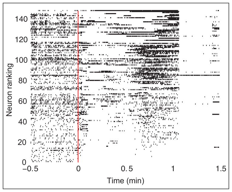
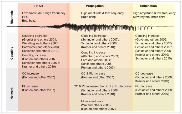
The brain is naturally considered as a network of interacting elements which, when functioning properly, produces an enormous range of dynamic, adaptable behavior. However, when elements of this network fail, pathological changes ensue, including epilepsy, one of the most common brain disorders. This review examines some aspects of cortical network organization that distinguish epileptic cortex from normal brain as well as the dynamics of network activity before and during seizures, focusing primarily on focal seizures. The review is organized around four phases of the seizure: the interictal period, onset, propagation, and termination. For each phase, the authors discuss the most common rhythmic characteristics of macroscopic brain voltage activity and outline the observed functional network features. Although the characteristics of functional networks that support the epileptic seizure remain an area of active research, the prevailing trends point to a complex set of network dynamics between, before, and during seizures.


Conflict of interest statement

Declaration of Conflicting Interests

The author(s) declared no potential conflicts of interest with respect to the research, authorship, and/or publication of this article.

Figures

Figure 1
Figure 1
Example ECoG recording. (A) An 8 × 8 electrode grid (1 cm spacing between electrodes) is placed directly on the cortical surface and recordings are made usually for several days or even weeks. The voltage data (B) recorded continuously for multiple days typically exhibit complicated dynamic activity.
Figure 2
Figure 2
Structural networks (left) represent physical connections between nodes (e.g., axons or white matter tracks). Here the nodes represent macroscopic brain regions that generate population voltage activity (middle). From the coupling between the node dynamics, functional networks are inferred (right) whose structure depends upon the choice of coupling measure and threshold.
Figure 3
Figure 3
Examples of three networks structures: regular (A), small-world (B), and random (C). In the regular network, a path between two nodes (green and yellow) is shown in green. For each network, the approximate average path length and clustering coefficient are shown.
Figure 4
Figure 4
Example of ictal dynamics. (A) Voltage trace from a single ECoG electrode during seizure. Visual inspection begins to suggest the different dynamic regimes. (B) A time–frequency spectrum of the voltage signal in (A). Warm (cool) colors indicate high (low) amplitude oscillations. As time evolves, the dominant rhythms slow.
Figure 5
Figure 5
Neuronal spike raster recorded in vivo from a human subject during seizure. The neurons (n = 149) are arranged according to increasing mean spike rate. The seizure begins at t = 0 min (solid red line) and ends near t = 1 min. Visual inspection suggests a nearly simultaneous cessation of spiking activity for most neurons at seizure termination. Adapted from Truccolo and others (2011).
Figure 6
Figure 6
Summary of functional network organization during different seizure stages. Three different stages of seizure—onset, propagation, and termination—are shown with a typical voltage trace during seizure. The characteristics of the rhythms (top row), coupling (middle row), and networks (bottom row) are indicated as observed in each state and with references to the literature. HFO = high-frequency oscillation, CC = clustering coefficient, PL = path length.

References

    1. Alarcon G, Binnie CD, Elwes RD, Polkey CE. Power spectrum and intracranial EEG patterns at seizure onset in partial epilepsy. Electroencephalogr Clin Neurophysiol. 1995;94(5):326–37. - PubMed
    1. Altenburg J, Vermeulen RJ, Strijers RLM, Fetter WPF, Stam CJ. Seizure detection in the neonatal EEG with synchronization likelihood. Clin Neurophysiol. 2003;114(1):50–5. - PubMed
    1. Ansari-Asl K, Senhadji L, Bellanger J-J, Wendling F. Quantitative evaluation of linear and nonlinear methods characterizing interdependencies between brain signals. Phys Rev E. 2006;74(3 pt 1):031916. - PMC - PubMed
    1. Antiqueira L, Rodrigues FA, van Wijk BCM, da F Costa L, Daffertshofer A. Estimating complex cortical networks via surface recordings—a critical note. Neuroimage. 2010;53(2):439–49. - PubMed
    1. Arnhold J, Grassberger P, Lehnertz K, Elger C. A robust method for detecting interdependences: application to intracranially recorded EEG. Physica D. 1999;134(4):419–30.

Publication types